
Search Count: 60,200
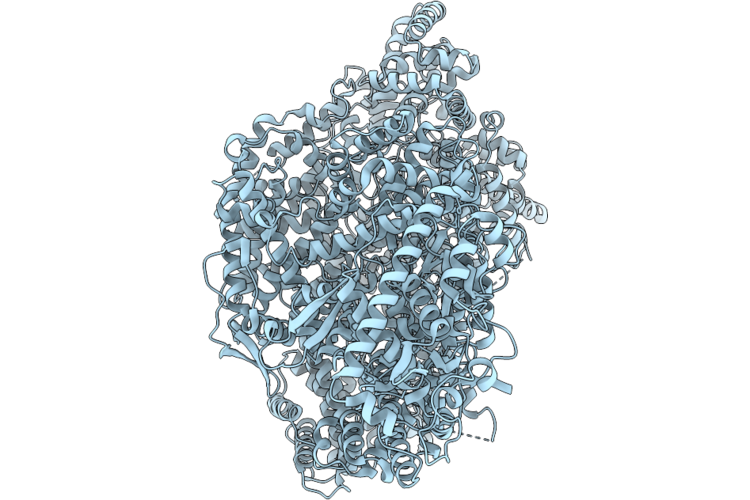 |
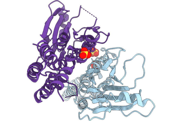
Cryo-Em Structure Of The Bacteriophage N4 Virion Rna Polymerase (Open Plug State)
Organism: Escherichia phage n4
Method: ELECTRON MICROSCOPY Resolution:3.35 Å Release Date: 2026-06-24 Classification: VIRAL PROTEIN |
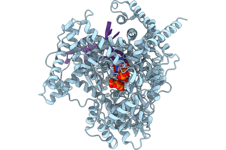 |
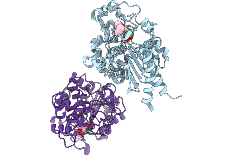
Cryo-Em Structure Of The Bacteriophage N4 Virion Rna Polymerase (Transcription Initiation Complex)
Organism: Escherichia phage n4, Synthetic construct
Method: ELECTRON MICROSCOPY Resolution:2.82 Å Release Date: 2026-06-24 Classification: VIRAL PROTEIN Ligands: GTP, MG |
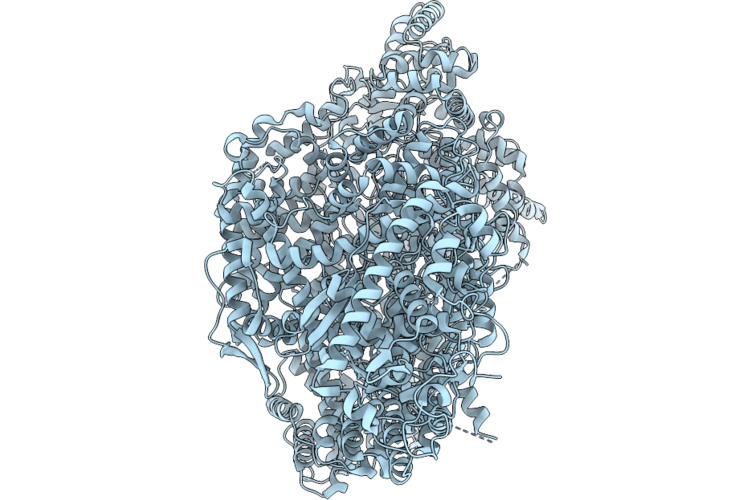 |
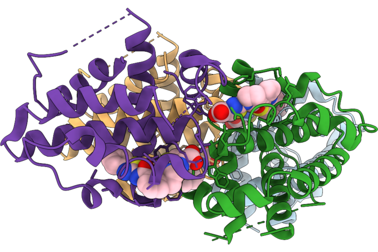
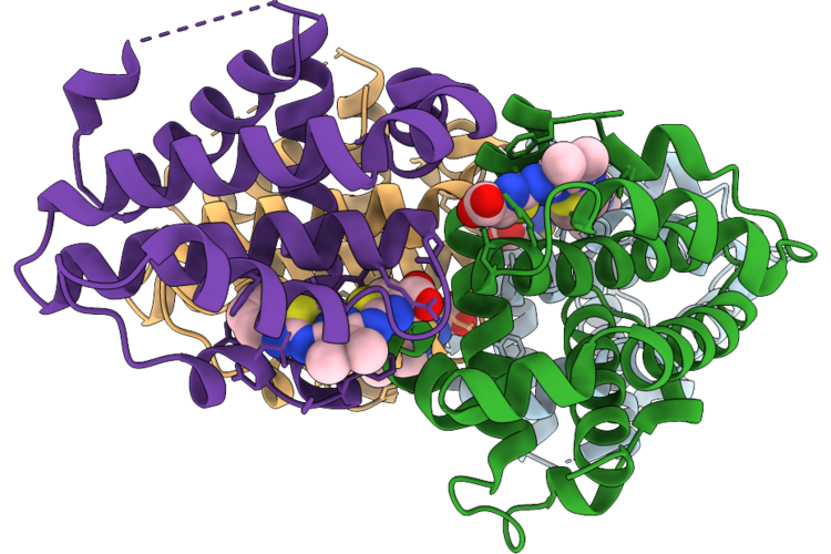
Cryo-Em Structure Of The Bacteriophage N4 Virion Rna Polymerase (Closed Plug State)
Organism: Escherichia phage n4
Method: ELECTRON MICROSCOPY Resolution:3.43 Å Release Date: 2026-06-24 Classification: VIRAL PROTEIN |
 |
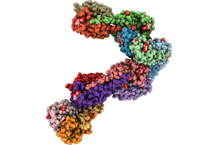
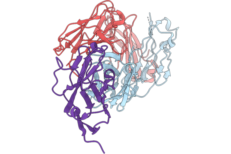
Sars-Cov-2 Rna-Dependent Rna Polymerase In Complex With 4'-Fla Nucleotide Analogue
Organism: Severe acute respiratory syndrome coronavirus 2
Method: ELECTRON MICROSCOPY Release Date: 2026-06-24 Classification: VIRAL PROTEIN/DNA Ligands: ZN, POP, A1DCZ, MG |
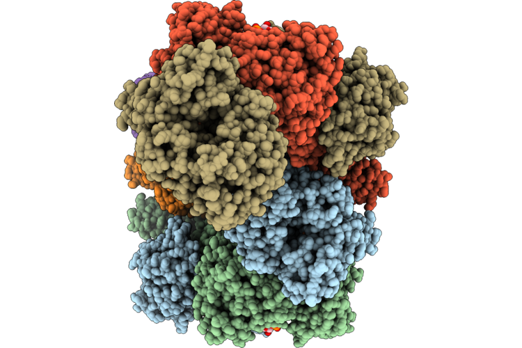 |
Cryoem Structure Of Adhe Spirosome From Clostridium Thermocellum Uncovered By Visual Proteomics.
Organism: Acetivibrio thermocellus dsm 1313
Method: ELECTRON MICROSCOPY Release Date: 2026-06-24 Classification: OXIDOREDUCTASE Ligands: FE, NAD |
 |
Crystal Structure Of The De Novo Designed Miniprotein Binder Against Pdl1, Gpx41.
Organism: Synthetic construct
Method: X-RAY DIFFRACTION Resolution:1.75 Å Release Date: 2026-06-24 Classification: DE NOVO PROTEIN |
 |
Organism: Xenorhabdus nematophila
Method: X-RAY DIFFRACTION Resolution:1.70 Å Release Date: 2026-06-24 Classification: SUGAR BINDING PROTEIN Ligands: EDO, CA, MG, SO4, PEG, BGC, PGE, CL, NA, 1PE |
 |
Crystal Structure Of The Substrate-Binding Domain Of S. Cerevisiae Ssc1 In Complex With Peptide Lslppvklhc
Organism: Saccharomyces cerevisiae s288c
Method: X-RAY DIFFRACTION Resolution:2.23 Å Release Date: 2026-06-24 Classification: CHAPERONE |
 |
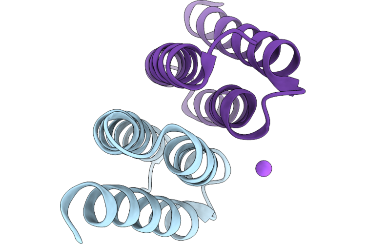
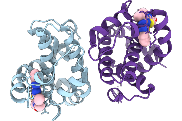
Organism: Homo sapiens
Method: X-RAY DIFFRACTION Resolution:2.49 Å Release Date: 2026-06-24 Classification: TRANSCRIPTION Ligands: SO4 |
 |
Crystal Structure Of Pdc-3 Beta-Lactamase Complexed With Boronic Acid Inhibitor Z2242032529
Organism: Pseudomonas aeruginosa
Method: X-RAY DIFFRACTION Resolution:1.98 Å Release Date: 2026-06-24 Classification: HYDROLASE/INHIBITOR Ligands: A1BI9 |
 |
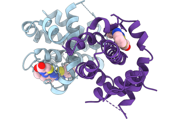
Organism: Homo sapiens
Method: X-RAY DIFFRACTION Resolution:1.70 Å Release Date: 2026-06-24 Classification: APOPTOSIS Ligands: A1I1S |
 |
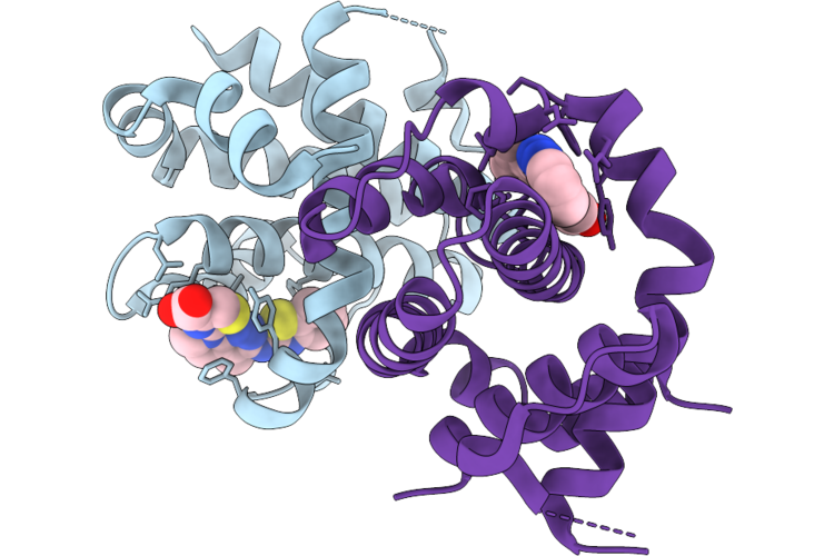
Organism: Homo sapiens
Method: X-RAY DIFFRACTION Resolution:3.34 Å Release Date: 2026-06-24 Classification: APOPTOSIS Ligands: A1I4A |
 |
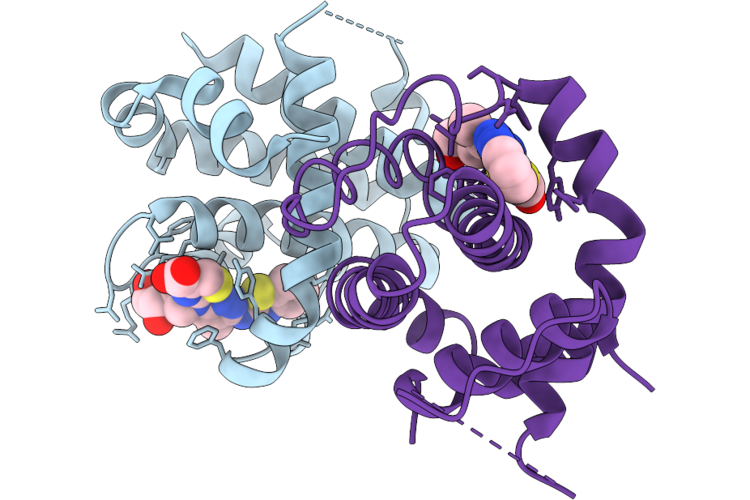
Organism: Homo sapiens
Method: X-RAY DIFFRACTION Resolution:2.10 Å Release Date: 2026-06-24 Classification: APOPTOSIS Ligands: A1I35 |
 |
Organism: Homo sapiens
Method: X-RAY DIFFRACTION Resolution:2.40 Å Release Date: 2026-06-24 Classification: APOPTOSIS Ligands: A1I37, CL |
 |
Organism: Homo sapiens
Method: X-RAY DIFFRACTION Resolution:1.80 Å Release Date: 2026-06-24 Classification: APOPTOSIS Ligands: A1I38, CL |
 |
Organism: Homo sapiens
Method: X-RAY DIFFRACTION Resolution:2.70 Å Release Date: 2026-06-24 Classification: APOPTOSIS Ligands: A1I39 |
 |
Organism: Homo sapiens
Method: X-RAY DIFFRACTION Resolution:1.50 Å Release Date: 2026-06-24 Classification: APOPTOSIS Ligands: A1I4B |
 |
Organism: Homo sapiens
Method: X-RAY DIFFRACTION Resolution:1.95 Å Release Date: 2026-06-24 Classification: APOPTOSIS Ligands: A1I36, A1I4C, CL |
 |
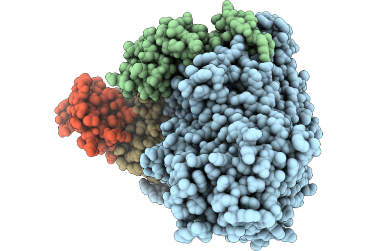
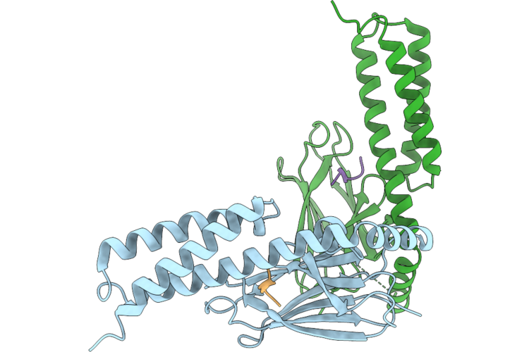
Organism: Homo sapiens
Method: ELECTRON MICROSCOPY Release Date: 2026-06-24 Classification: SIGNALING PROTEIN/IMMUNE SYSTEM |
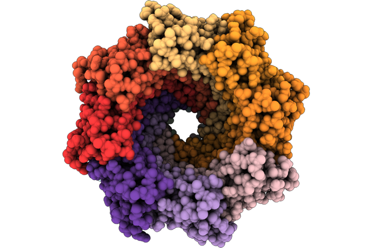 |
C1 Symmetry Cryoem Structure Of The Soluble-Wraped Membranous Portion Of Mspa (Mycobacterium Smegmatis Porin), Dimerized Along The Native Interface.
Organism: Mycolicibacterium smegmatis mc2 155
Method: ELECTRON MICROSCOPY Resolution:3.34 Å Release Date: 2026-06-24 Classification: DE NOVO PROTEIN |