

Search Count: 16,206
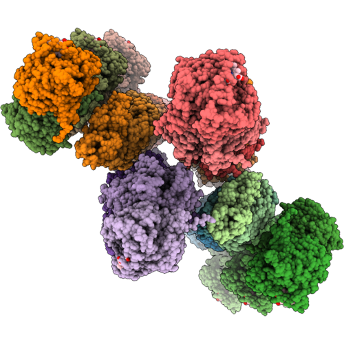 |
Structure Of Stacked Allo-Activated Cbs Conformer By Allosteric Sam - By Helical Approach
Organism: Homo sapiens
Method: ELECTRON MICROSCOPY Resolution:8.50 Å Release Date: 2026-11-04 Classification: LYASE Ligands: HEM Modified Residue(s): LLP |
Organism: Homo sapiens
Method: ELECTRON MICROSCOPY
Release Date: 2026-11-04
Ligands: HEM
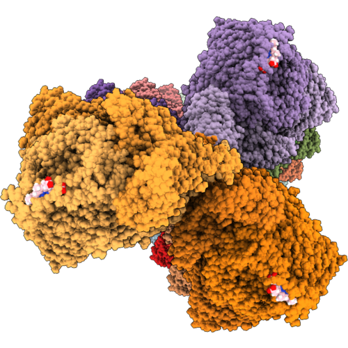 |
Structure Of Trans-Basal Conformer Of Human Cbs Encompassing 16-18 Dimer Repeating Units (In Absence Of Substrate And Allosteric Ligand)- By Helical Approach
Organism: Homo sapiens
Method: ELECTRON MICROSCOPY Release Date: 2026-11-04 Classification: LYASE Ligands: HEM Modified Residue(s): LLP |
Organism: Homo sapiens
Method: ELECTRON MICROSCOPY
Release Date: 2026-11-04
Ligands: HEM
 |
Ethanolamine Ammonia-Lyase, Adocbl/Substrate-Free
Organism: Escherichia coli (strain k12)
Method: X-RAY DIFFRACTION Resolution:2.00 Å Release Date: 2018-09-19 Classification: LYASE Ligands: 5AD, GOL, B12 |
Organism: Escherichia coli (strain k12)
Method: X-RAY DIFFRACTION
Release Date: 2018-09-19
Ligands: 5AD, GOL, B12
 |
Ethanolamine Ammonia-Lyase, Adocbl/2-Amino-1-Propanol
Organism: Escherichia coli k-12
Method: X-RAY DIFFRACTION Resolution:2.05 Å Release Date: 2018-09-19 Classification: LYASE Ligands: 5AD, GOL, B12 |
Organism: Escherichia coli k-12
Method: X-RAY DIFFRACTION
Release Date: 2018-09-19
Ligands: 5AD, GOL, B12
 |
Ethanolamine Ammonia-Lyase Complexed With Adomecbl
Organism: Escherichia coli
Method: X-RAY DIFFRACTION Resolution:2.13 Å Release Date: 2023-03-29 Classification: LYASE Ligands: GOL, NH4, FWK, B12 |
Organism: Escherichia coli
Method: X-RAY DIFFRACTION
Release Date: 2023-03-29
Ligands: GOL, NH4, FWK, B12
 |
Ethanolamine Ammonia-Lyase Complexed With Adomecbl In The Presence Of Substrate
Organism: Escherichia coli
Method: X-RAY DIFFRACTION Resolution:2.07 Å Release Date: 2023-03-29 Classification: LYASE Ligands: FWK, GOL, NH4, B12 |
Organism: Escherichia coli
Method: X-RAY DIFFRACTION
Release Date: 2023-03-29
Ligands: FWK, GOL, NH4, B12
 |
Crystal Structure Of Multi-Functional Polysaccharide Lyase Smlt1473-H168A From Stenotrophomonas Maltophilia (Strain K279A) At Ph-5.0
Organism: Stenotrophomonas maltophilia k279a
Method: X-RAY DIFFRACTION Resolution:2.60 Å Release Date: 2021-10-27 Classification: LYASE Ligands: PEG, SO4 |
Organism: Stenotrophomonas maltophilia k279a
Method: X-RAY DIFFRACTION
Release Date: 2021-10-27
Ligands: PEG, SO4
 |
Crystal Structure Of Multi-Functional Polysaccharide Lyase Smlt1473 From Stenotrophomonas Maltophilia (Strain K279A) In Apo Form At Ph 5.5
Organism: Stenotrophomonas maltophilia k279a
Method: X-RAY DIFFRACTION Resolution:2.06 Å Release Date: 2021-10-27 Classification: LYASE |
Organism: Stenotrophomonas maltophilia k279a
Method: X-RAY DIFFRACTION
Release Date: 2021-10-27
 |
Crystal Structure Of Multi-Functional Polysaccharide Lyase Smlt1473 From Stenotrophomonas Maltophilia (Strain K279A) In Apo Form At Ph 8.5
Organism: Stenotrophomonas maltophilia k279a
Method: X-RAY DIFFRACTION Resolution:2.43 Å Release Date: 2021-10-27 Classification: LYASE Ligands: SO4 |
Organism: Stenotrophomonas maltophilia k279a
Method: X-RAY DIFFRACTION
Release Date: 2021-10-27
Ligands: SO4
 |
Crystal Structure Of Multi-Functional Polysaccharide Lyase Smlt1473 (Wt) From Stenotrophomonas Maltophilia (Strain K279A) In Apo Form At Ph 9.0
Organism: Stenotrophomonas maltophilia k279a
Method: X-RAY DIFFRACTION Resolution:2.50 Å Release Date: 2021-10-27 Classification: LYASE Ligands: SO4 |
Organism: Stenotrophomonas maltophilia k279a
Method: X-RAY DIFFRACTION
Release Date: 2021-10-27
Ligands: SO4
 |
Crystal Structure Of Multi-Functional Polysaccharide Lyase Smlt1473 (Wt) From Stenotrophomonas Maltophilia (Strain K279A) In Apo Form At Ph 6.5
Organism: Stenotrophomonas maltophilia k279a
Method: X-RAY DIFFRACTION Resolution:2.28 Å Release Date: 2021-10-27 Classification: LYASE Ligands: SO4, PEG |
Organism: Stenotrophomonas maltophilia k279a
Method: X-RAY DIFFRACTION
Release Date: 2021-10-27
Ligands: SO4, PEG
 |
Crystal Structure Of Multi-Functional Polysaccharide Lyase Smlt1473 (Wt) From Stenotrophomonas Maltophilia (Strain K279A) In Apo Form At Ph 7.0
Organism: Stenotrophomonas maltophilia k279a
Method: X-RAY DIFFRACTION Resolution:2.20 Å Release Date: 2021-10-27 Classification: LYASE Ligands: SO4 |
Organism: Stenotrophomonas maltophilia k279a
Method: X-RAY DIFFRACTION
Release Date: 2021-10-27
Ligands: SO4
 |
Crystal Structure Of Multi-Functional Polysaccharide Lyase Smlt1473 (Wt) From Stenotrophomonas Maltophilia (Strain K279A) In Apo Form At Ph 5.0
Organism: Stenotrophomonas maltophilia k279a
Method: X-RAY DIFFRACTION Resolution:2.63 Å Release Date: 2021-10-27 Classification: LYASE Ligands: SO4, PEG |
Organism: Stenotrophomonas maltophilia k279a
Method: X-RAY DIFFRACTION
Release Date: 2021-10-27
Ligands: SO4, PEG
 |
Crystal Structure Of Multi-Functional Polysaccharide Lyase Smlt1473 (Wt) From Stenotrophomonas Maltophilia (Strain K279A) In Mana Bound Form At Ph-7.0
Organism: Stenotrophomonas maltophilia k279a
Method: X-RAY DIFFRACTION Resolution:2.43 Å Release Date: 2021-10-27 Classification: LYASE Ligands: SO4 |
Organism: Stenotrophomonas maltophilia k279a
Method: X-RAY DIFFRACTION
Release Date: 2021-10-27
Ligands: SO4
 |
Crystal Structure Of Multi-Functional Polysaccharide Lyase Smlt1473 (Wt) From Stenotrophomonas Maltophilia (Strain K279A) In Mana Bound Form At Ph-5.0
Organism: Stenotrophomonas maltophilia k279a
Method: X-RAY DIFFRACTION Resolution:2.31 Å Release Date: 2021-10-27 Classification: LYASE Ligands: PEG, SO4 |
Organism: Stenotrophomonas maltophilia k279a
Method: X-RAY DIFFRACTION
Release Date: 2021-10-27
Ligands: PEG, SO4
 |
Crystal Structure Of Ethanolamine Ammonia-Lyase From Escherichia Coli Complexed With Adeninylpentylcobalamin And Ethanolamine
Organism: Escherichia coli
Method: X-RAY DIFFRACTION Resolution:2.25 Å Release Date: 2010-06-02 Classification: LYASE Ligands: COY, ETA, GOL, SO4 |
Organism: Escherichia coli
Method: X-RAY DIFFRACTION
Release Date: 2010-06-02
Ligands: COY, ETA, GOL, SO4
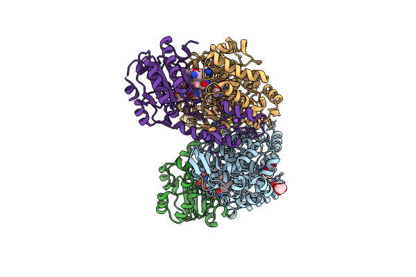 |
Crystal Structure Of Ethanolamine Ammonia-Lyase From Escherichia Coli Complexed With Cn-Cbl And Ethanolamine
Organism: Escherichia coli
Method: X-RAY DIFFRACTION Resolution:2.10 Å Release Date: 2010-06-02 Classification: LYASE Ligands: ETA, GOL, B12, NA |
Organism: Escherichia coli
Method: X-RAY DIFFRACTION
Release Date: 2010-06-02
Ligands: ETA, GOL, B12, NA
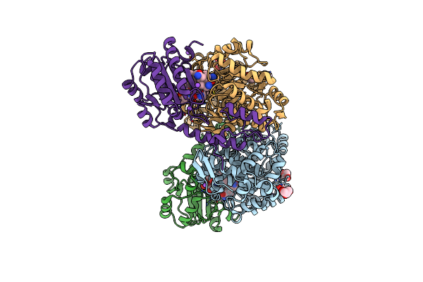 |
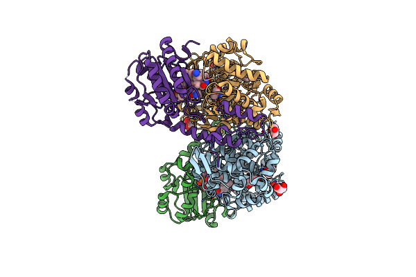
Crystal Structure Of Ethanolamine Ammonia-Lyase From Escherichia Coli Complexed With Cn-Cbl (Substrate-Free Form)
Organism: Escherichia coli
Method: X-RAY DIFFRACTION Resolution:2.10 Å Release Date: 2010-06-02 Classification: LYASE Ligands: GOL, NA, B12 |
Organism: Escherichia coli
Method: X-RAY DIFFRACTION
Release Date: 2010-06-02
Ligands: GOL, NA, B12
 |
Crystal Structure Of Ethanolamine Ammonia-Lyase From Escherichia Coli Complexed With Cn-Cbl And 2-Amino-1-Propanol
Organism: Escherichia coli
Method: X-RAY DIFFRACTION Resolution:2.05 Å Release Date: 2010-06-02 Classification: LYASE Ligands: 2A1, GOL, B12 |
Organism: Escherichia coli
Method: X-RAY DIFFRACTION
Release Date: 2010-06-02
Ligands: 2A1, GOL, B12
 |
Crystal Structure Of Ethanolamine Ammonia-Lyase From Escherichia Coli Complexed With Cn-Cbl And (R)-2-Amino-1-Propanol
Organism: Escherichia coli
Method: X-RAY DIFFRACTION Resolution:2.10 Å Release Date: 2011-08-03 Classification: LYASE Ligands: 2A3, B12 |
Organism: Escherichia coli
Method: X-RAY DIFFRACTION
Release Date: 2011-08-03
Ligands: 2A3, B12