

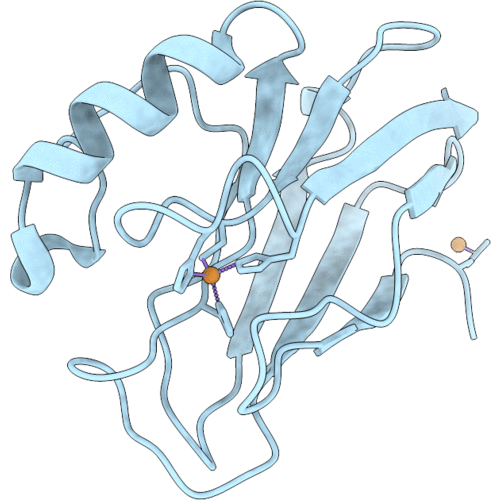
9YZ6 image
Deposition Date 2025-10-30
Release Date 2026-09-09
Last Version Date 2026-09-09
Entry Detail
PDB ID:
9YZ6
Title:
Pseudomonas aeruginosa azurin mutant - M121H
Biological Source:
Source Organism(s):
Expression System(s):
Method Details:
Experimental Method:
Resolution:
2.47 Å
R-Value Free:
0.33
R-Value Work:
0.27
R-Value Observed:
0.28
Space Group:
C 2 2 21
Macromolecular Entities
Polymer Type:polypeptide(L)
Molecule:Azurin
Gene (Uniprot):azu
Mutagens:M121H
Chain IDs:A
Chain Length:128
Number of Molecules:1
Biological Source:Pseudomonas aeruginosa
Ligand Molecules
Primary Citation
Structural basis for organism-dependent effects on spectroscopic properties of type 1 copper azurin by the same axial methionine to histidine mutation.
J.Inorg.Biochem. 277 113209 113209 (2026)
PMID: 41518912 DOI: 10.1016/j.jinorgbio.2025.113209

Abstact

Cupredoxins are a family of copper proteins that mediate electron transfer in key biological processes, and understanding structural features responsible for their spectroscopic properties is important for elucidating structure-function relationships. Here, we characterize the effect of mutation of the axial methionine to histidine in azurin from Pseudomonas aeruginosa (M121HAz-PA) using UV-Vis absorption, electron paramagnetic resonance (EPR) spectroscopy, and X-ray crystallography to examine how the same M121H mutation affects spectroscopic properties differently from its homolog in Alcaligenes denitrificans (M121HAz-AD). M121HAz-PA shows a pH-dependent UV-vis spectral change from pH 4 to pH 8, with unusual EPR features, indicating changes in coordination electronic structure. Azide binding titrations produce EPR spectra intermediate between type 1 copper (T1Cu) and type 2 copper (T2Cu), supporting that changes of (exogenous) ligand coordination drive significant geometric and electronic rearrangements. X-ray crystallography reveals differences in axial His121 coordination mode to Cu(II) and its orientation between M121HAz-PA and M121HAz-AD, providing a structural basis for the observed spectroscopic differences. These findings provide insights into how subtle variations in ligand coordination affect the electronic structure in Cu-binding sites, offering a basis for the rational design of metalloproteins.

Legend

Protein

Chemical

Disease

Primary Citation of related structures
Feedback Form
Name
Email
Institute
Feedback