

9YK1 image
Deposition Date 2025-10-06
Release Date 2026-03-18
Last Version Date 2026-04-01
Entry Detail
PDB ID:
9YK1
Title:
Room-temperature X-ray structure of D132N Bacillus halodurans RNase H1 in complex with RNA/DNA duplex
Biological Source:
Source Organism(s):
Expression System(s):
Method Details:
Experimental Method:
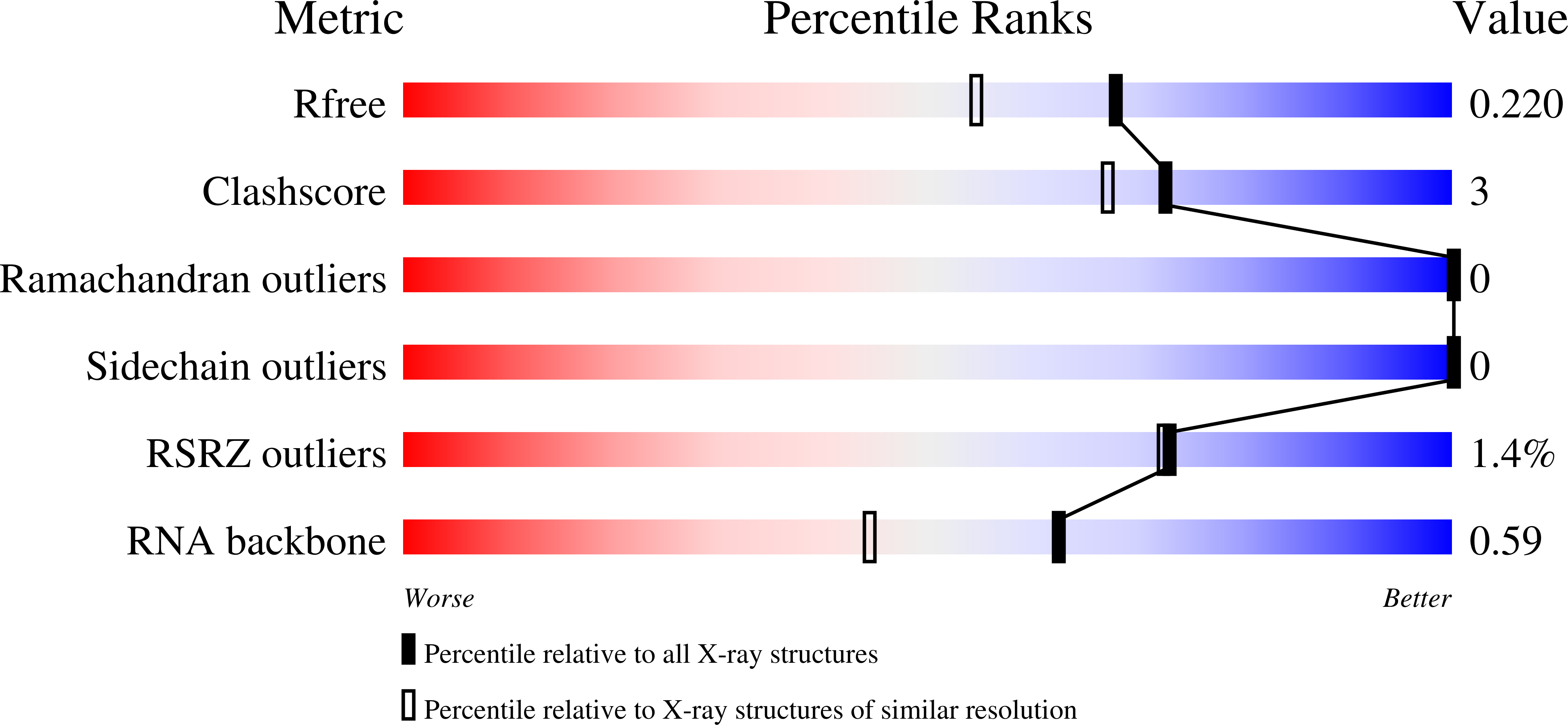
Resolution:
1.80 Å
R-Value Free:
0.21
R-Value Work:
0.18
R-Value Observed:
0.18
Space Group:
C 1 2 1
Macromolecular Entities
Structures with similar UniProt ID
Protein Blast
Polymer Type:polypeptide(L)
Molecule:Ribonuclease H
Gene (Uniprot):rnhA
Chain IDs:A
Chain Length:139
Number of Molecules:1
Biological Source:Halalkalibacterium halodurans
Polymer Type:polyribonucleotide
Molecule:RNA (5'-R(*UP*CP*GP*AP*CP*A)-
Chain IDs:B
Chain Length:6
Number of Molecules:1
Biological Source:Halalkalibacterium halodurans
Polymer Type:polydeoxyribonucleotide
Molecule:DNA (5'-D(*AP*TP*GP*TP*CP*G)-
Chain IDs:C
Chain Length:6
Number of Molecules:1
Biological Source:Halalkalibacterium halodurans
Primary Citation
Structural insights into RNase H catalytic mechanism from room-temperature X-ray and neutron crystallography of apo- and RNA/DNA hybrid-bound enzyme.
Curr Res Struct Biol 11 100188 100188 (2026)
PMID: 41853683 DOI: 10.1016/j.crstbi.2026.100188

Abstact

RNase H enzymes are sequence-nonspecific endonucleases that cleave RNA strands in RNA/DNA hybrid duplexes, an enzymatic process essential in DNA replication and repair in both prokaryotes and eukaryotes. Also, RNase H activity of the reverse transcriptase in human immunodeficiency viruses (HIV-1 and HIV-2) is indispensable for the viral replication cycle. RNase H enzymes play an central role in the development of gene therapies and are targets for novel antivirals. It is therefore of great importance to gain a detailed understanding of the RNase H catalytic mechanism to improve drug design. We utilized Bacillus halodurans RNase H1 (BhRNase H1) to shed light on its function and catalytic mechanism. Room-temperature neutron crystallography of the wild-type and inactive D132N mutant enzymes revealed that E109, belonging to the catalytic DEDD motif, can change its protonation state, allowing us to propose its role in the protonation of the leaving O3' hydroxyl group of RNA. X-ray crystallography has demonstrated the ability of the RNA/DNA duplex to slide along the protein surface upon metal ion binding at site M(A), transforming a product mimic into a Michaelis-like complex, which confirms an essential role of the M(A) metal ion in catalysis.

Legend

Protein

Chemical

Disease

Primary Citation of related structures
Feedback Form
Name
Email
Institute
Feedback