

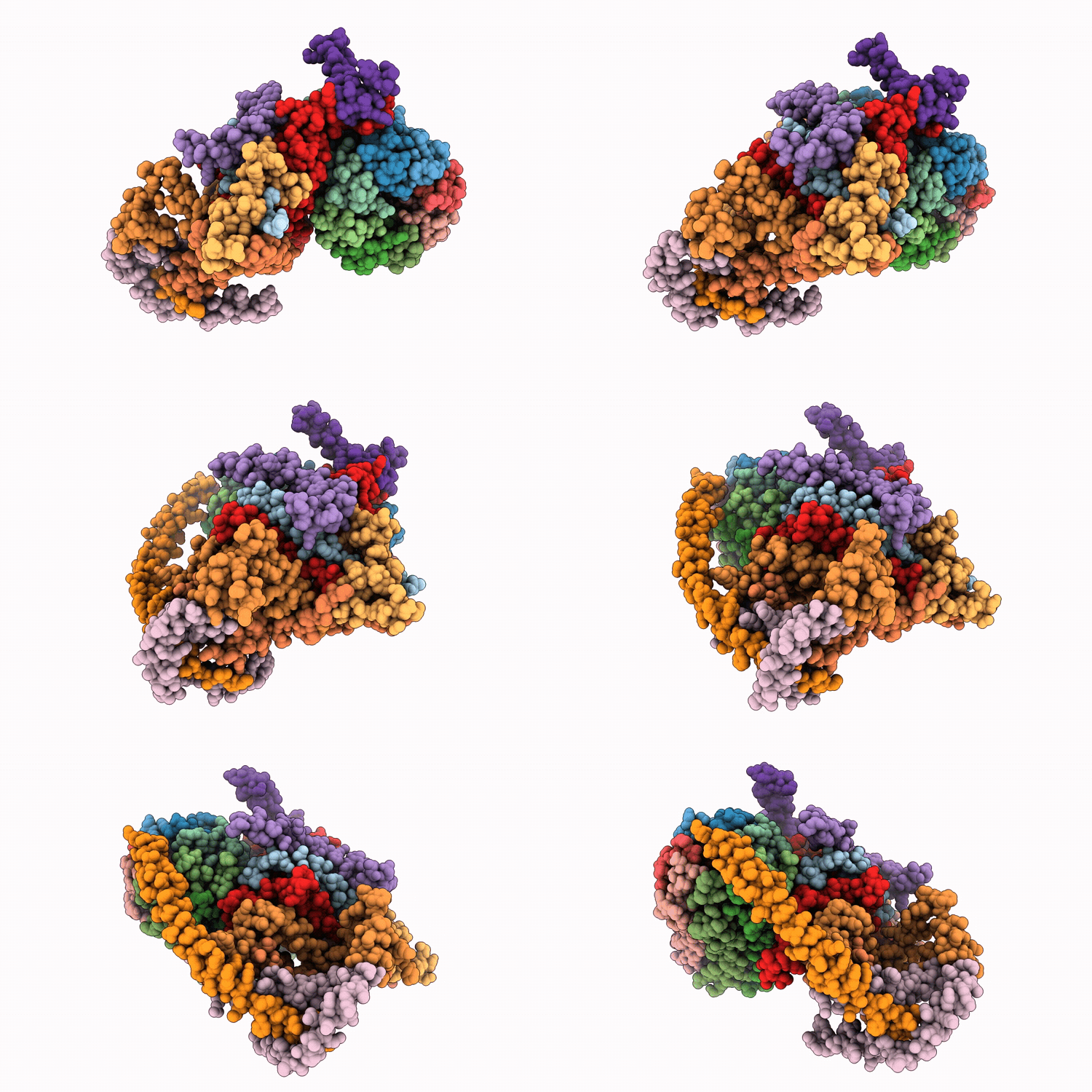
9W2T image
Deposition Date 2025-07-28
Release Date 2026-04-01
Last Version Date 2026-05-20
Entry Detail
PDB ID:
9W2T
Keywords:
Title:
Cryo-EM structure of Fo domain of FoF1-ATPase monomer state on the bovine heart submitochondrial particles
Biological Source:
Source Organism(s):
Bos taurus (Taxon ID: 9913)
Method Details:
Experimental Method:
Resolution:
4.10 Å
Aggregation State:
PARTICLE
Reconstruction Method:
SINGLE PARTICLE
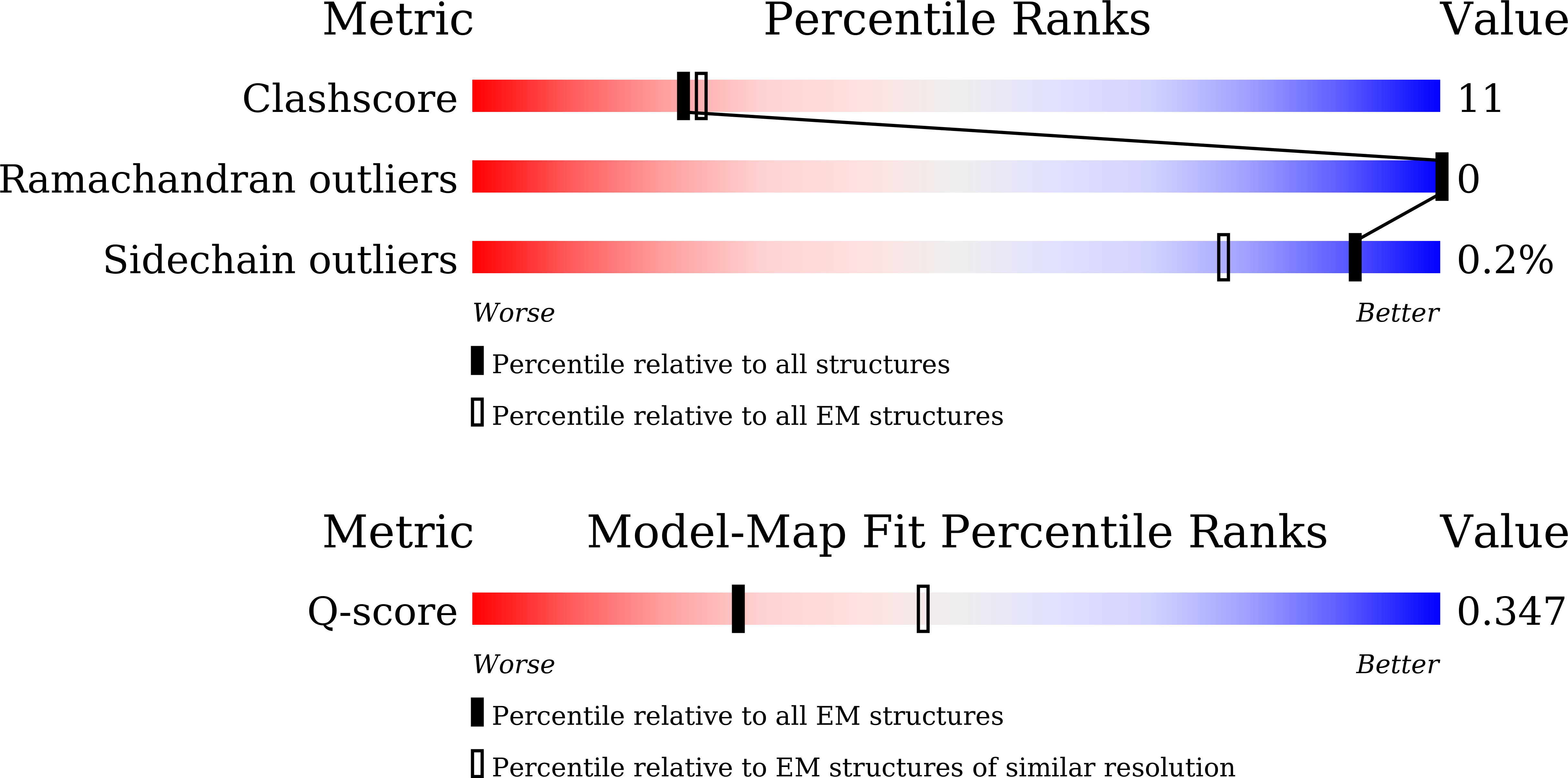
Macromolecular Entities
Structures with similar UniProt ID
Protein Blast
Polymer Type:polypeptide(L)
Molecule:ATP synthase F(0) complex sub
Gene (Uniprot):MT-ATP8
Chain IDs:A (auth: 8)
Chain Length:41
Number of Molecules:1
Biological Source:Bos taurus
Structures with similar UniProt ID
Protein Blast
Polymer Type:polypeptide(L)
Molecule:ATP synthase F(0) complex sub
Gene (Uniprot):ATP5MC1
Chain IDs:B (auth: K), C (auth: L), D (auth: M), E (auth: N), F (auth: O), G (auth: P), H (auth: Q), I (auth: R)
Chain Length:75
Number of Molecules:8
Biological Source:Bos taurus
Structures with similar UniProt ID
Protein Blast
Polymer Type:polypeptide(L)
Molecule:ATP synthase F(0) complex sub
Gene (Uniprot):MT-ATP6
Chain IDs:J (auth: a)
Chain Length:224
Number of Molecules:1
Biological Source:Bos taurus
Structures with similar UniProt ID
Protein Blast
Polymer Type:polypeptide(L)
Molecule:ATP synthase peripheral stalk
Gene (Uniprot):ATP5PB
Chain IDs:K (auth: b)
Chain Length:112
Number of Molecules:1
Biological Source:Bos taurus
Structures with similar UniProt ID
Protein Blast
Polymer Type:polypeptide(L)
Molecule:ATP synthase peripheral stalk
Gene (Uniprot):ATP5PD
Chain IDs:L (auth: d)
Chain Length:51
Number of Molecules:1
Biological Source:Bos taurus
Structures with similar UniProt ID
Protein Blast
Polymer Type:polypeptide(L)
Molecule:ATP synthase F(0) complex sub
Gene (Uniprot):ATP5ME
Chain IDs:M (auth: e)
Chain Length:69
Number of Molecules:1
Biological Source:Bos taurus
Structures with similar UniProt ID
Protein Blast
Polymer Type:polypeptide(L)
Molecule:ATP synthase F(0) complex sub
Gene (Uniprot):ATP5MF
Chain IDs:N (auth: f)
Chain Length:83
Number of Molecules:1
Biological Source:Bos taurus
Structures with similar UniProt ID
Protein Blast
Polymer Type:polypeptide(L)
Molecule:ATP synthase F(0) complex sub
Gene (Uniprot):ATP5MG
Chain IDs:O (auth: g)
Chain Length:79
Number of Molecules:1
Biological Source:Bos taurus
Structures with similar UniProt ID
Protein Blast
Polymer Type:polypeptide(L)
Molecule:ATP synthase F(0) complex sub
Gene (Uniprot):ATP5MJ
Chain IDs:P (auth: j)
Chain Length:48
Number of Molecules:1
Biological Source:Bos taurus
Structures with similar UniProt ID
Protein Blast
Polymer Type:polypeptide(L)
Molecule:ATP synthase F(0) complex sub
Gene (Uniprot):ATP5MK
Chain IDs:Q (auth: k)
Chain Length:36
Number of Molecules:1
Biological Source:Bos taurus
Modified Residue
Compound ID Chain ID Parent Comp ID Details 2D Image
M3L B LYS modified residue
Ligand Molecules
Primary Citation
Structures of respiratory supercomplexes and ATP synthase oligomers in mammalian mitochondrial inner membrane.
Nat Commun 17 ? ? (2026)
PMID: 41844608 DOI: 10.1038/s41467-026-70578-x

Abstact

Understanding the functional mechanisms of membrane protein complexes requires structural analysis within their native membrane environment. Here, we applied cryo-electron microscopy to determine the structures of F(o)F(1) ATP synthase and respiratory supercomplexes (SCs) on sub-mitochondrial particles (SMPs) isolated from bovine heart mitochondria. Most F(o)F(1) complexes were observed as dimers stabilized by the regulatory factor IF(1), and a tetrameric assembly comprising two F(o)F(1)-IF(1) dimers arranged linearly was also identified. This finding indicates that the tetrameric units of F(o)F(1) are present in the mitochondrial inner membrane and contribute to shaping cristae tips in mammalian mitochondria. F(o) domain maps resolve the e-subunit- c(8)-ring interface and show no discrete density for a tightly bound lipid within the c(8)-ring. In addition to the previously reported SCs compositions CI(1)CIII(2)CIV(1) and CI(1)CIII(2)CIV(2), our analysis identified an additional assembly with the composition CI(1)CIII(2)CIV(3), as well as a CI(2)CIII(2)CIV(6) mega-complex. This approach enables rapid structural determination of F(o)F(1) ATP synthase and SCs from minimal membrane fractions, providing a foundation for elucidating the molecular basis of metabolic disorders and mitochondrial diseases at the level of higher-order architecture.

Legend

Protein

Chemical

Disease

Primary Citation of related structures
Feedback Form
Name
Email
Institute
Feedback