

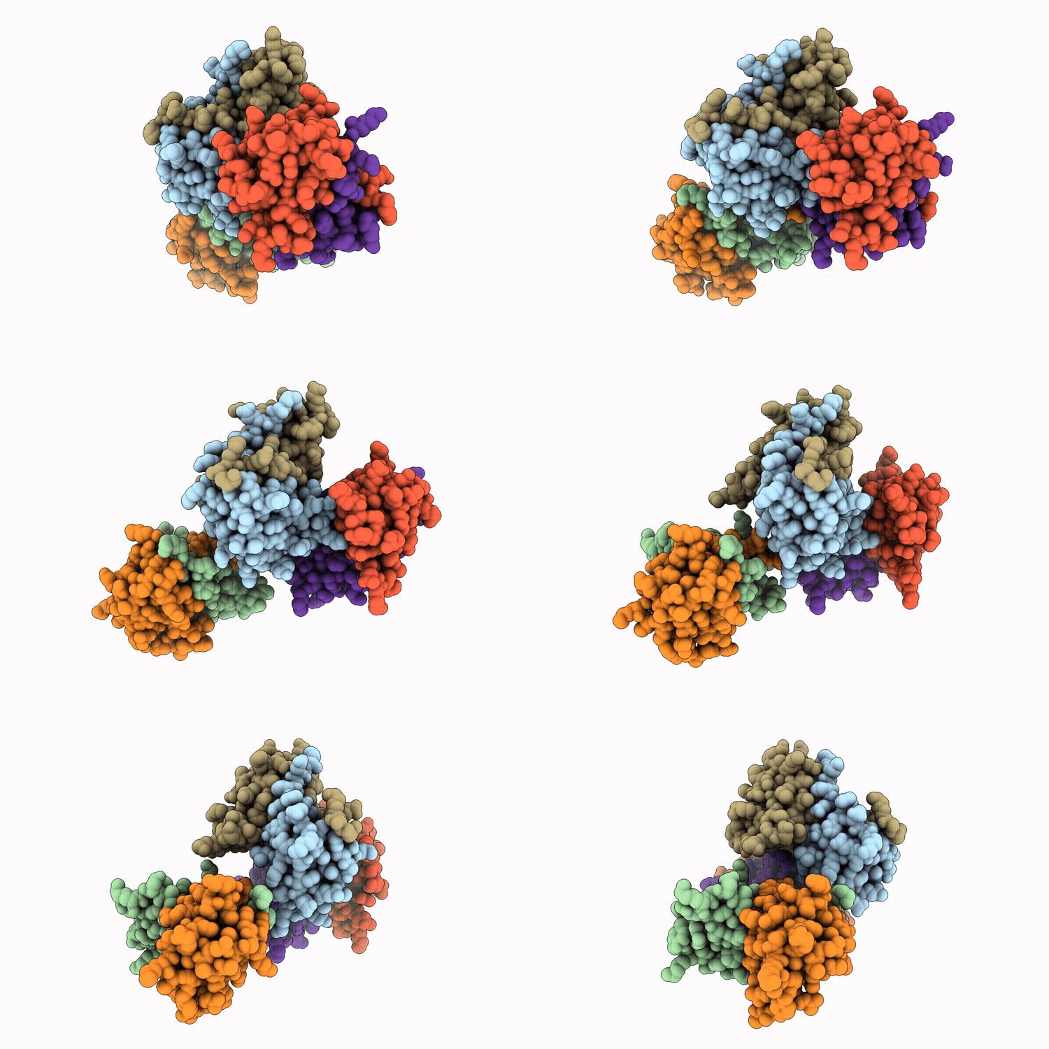
9VT3 image
Deposition Date 2025-07-09
Release Date 2026-05-27
Last Version Date 2026-05-27
Entry Detail
PDB ID:
9VT3
Title:
SeMet crystal structure of Aca3 from Neisseria meningitidis
Biological Source:
Source Organism(s):
Expression System(s):
Method Details:
Experimental Method:
Resolution:
2.09 Å
R-Value Free:
0.23
R-Value Work:
0.20
R-Value Observed:
0.20
Space Group:
P 21 21 21

Macromolecular Entities
Protein Blast
Polymer Type:polypeptide(L)
Molecule:Aca3
Chain IDs:A, B, C, D, E, F
Chain Length:76
Number of Molecules:6
Biological Source:Neisseria meningitidis
Primary Citation
Structural Basis of Recognition of Anti-CRISPR Operon by Aca3.
Faseb J. 40 e71512 e71512 (2026)
PMID: 41630633 DOI: 10.1096/fj.202502389RR

Abstact

The CRISPR-Cas system equips bacteria with adaptive immunity by storing fragments of invading nucleic acids in CRISPR loci and deploying Cas proteins to recognize and degrade matching sequences. In turn, bacteriophages have evolved small anti-CRISPR (Acr) that neutralize diverse CRISPR-Cas types. Acr genes are often co-encoded with transcriptional regulators called anti-CRISPR-associated (Aca) proteins, which suppress acr expression. Although 13 Aca families have been identified through bioinformatic analysis, detailed information on their target DNA-binding mechanisms and the inhibition of acr expression remains limited. Here, we report the high-resolution structure of Aca3 and delineate its DNA-binding interface. We demonstrate that Aca3 selectively recognizes inverted repeats upstream of its cognate acr gene, AcrIIC1. Mutational analyses of key helix-turn-helix residues confirm their essential roles in promoter engagement. Together, these results reveal the molecular basis for Aca3-mediated control of anti-CRISPR expression and expand our understanding of regulatory strategies that phages employ to modulate host CRISPR-Cas immunity.

Legend

Protein

Chemical

Disease

Primary Citation of related structures
Feedback Form
Name
Email
Institute
Feedback