9UYA image
Deposition Date 2025-05-15
Release Date 2026-05-13
Last Version Date 2026-05-13
Entry Detail
PDB ID:
9UYA
Keywords:
Title:
Crystal structure of phospholipase D form Streptomyces avermitilis
Biological Source:
Expression System(s):
Method Details:
Experimental Method:
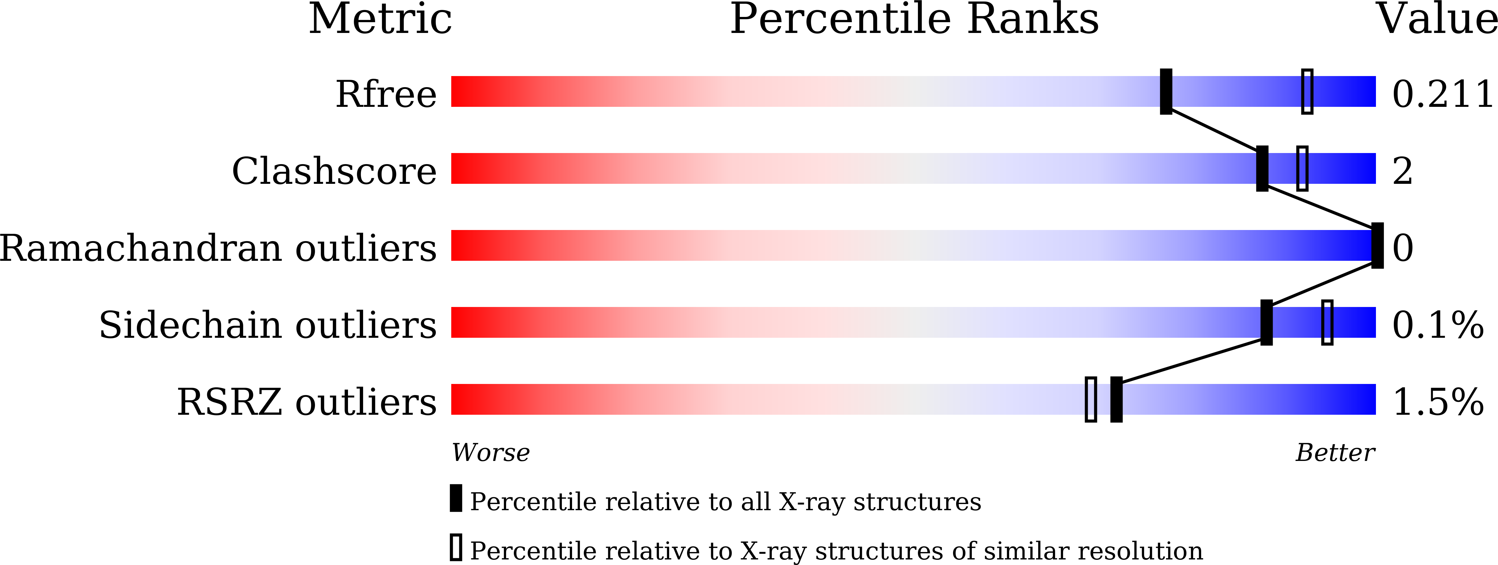
Resolution:
2.20 Å
R-Value Free:
0.21
R-Value Work:
0.17
R-Value Observed:
0.17
Space Group:
P 21 21 21
Macromolecular Entities
Structures with similar UniProt ID
Protein Blast
Polymer Type:polypeptide(L)
Molecule:Alkaline phosphatase, seceret
Gene (Uniprot):phoD1
Chain IDs:A, B, C, D
Chain Length:524
Number of Molecules:4
Biological Source:Streptomyces avermitilis MA-4680 = NBRC 14893
Primary Citation
x-Ray Structure of Streptomyces avermitilis Phospholipase D Reveals a Ca 2+ -Stabilized Expanded Active-Site Cleft Adapted for Phospholipid Binding.
Proteins ? ? ? (2026)
PMID: 42062768 DOI: 10.1002/prot.70142

Abstact

Phospholipase D (PLD) catalyzes the hydrolysis of phospholipids to generate phosphatidic acid and free head groups such as choline. Among bacterial PLD enzymes, Streptomyces chromofuscus PLD (SchPLD), a member of the alkaline phosphatase D (PhoD) superfamily, exhibits unique Ca(2+)-dependent phospholipase activity. Here, we determined the crystal structure of a PhoD-type PLD from S. avermitilis (SaPLD) at a 2.2-A resolution, which shares 86% sequence identity with SchPLD. The structure revealed the conserved Fe-Ca-Ca catalytic center characteristic of PhoD enzymes. In addition, we identified novel Ca(2+) binding sites surrounding the active site pocket. SaPLD exhibited negligible activity in the absence of Ca(2+) but showed strong activation in the presence of Ca(2+), consistent with previous observations for SchPLD. The overall structure of SaPLD lacks the C-terminal alpha-helix that covers the active site in Bacillus subtilis PhoD, resulting in an expanded hydrophobic cleft suited for bulky phospholipid substrates binding. Molecular dynamics modeling with phosphatidylcholine (PC) indicated that its two oleoyl chains fit well within this cleft, and that the choline head group is accommodated by a distinct cavity formed by Asn217, Leu346, and Asn357. This cavity geometry likely disfavors phosphatidylethanolamine or phosphatidylserine, explaining the preference for PC substrates. These findings provide the first structural insights into the Ca(2+)-stabilized expanded active site of a PhoD-type PLD and clarify the molecular basis for its phospholipid specificity.

Legend

Protein

Chemical

Disease

Primary Citation of related structures
Feedback Form
Name
Email
Institute
Feedback