9UNP image
Deposition Date 2025-04-24
Release Date 2026-02-18
Last Version Date 2026-05-13
Entry Detail
PDB ID:
9UNP
Title:
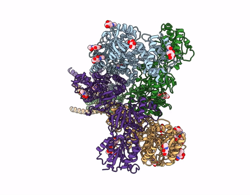
native NMDA receptor-GluN1/N2A/N2B-S2 in the closed state
Biological Source:
Source Organism(s):
Mus musculus (Taxon ID: 10090)
Method Details:
Experimental Method:
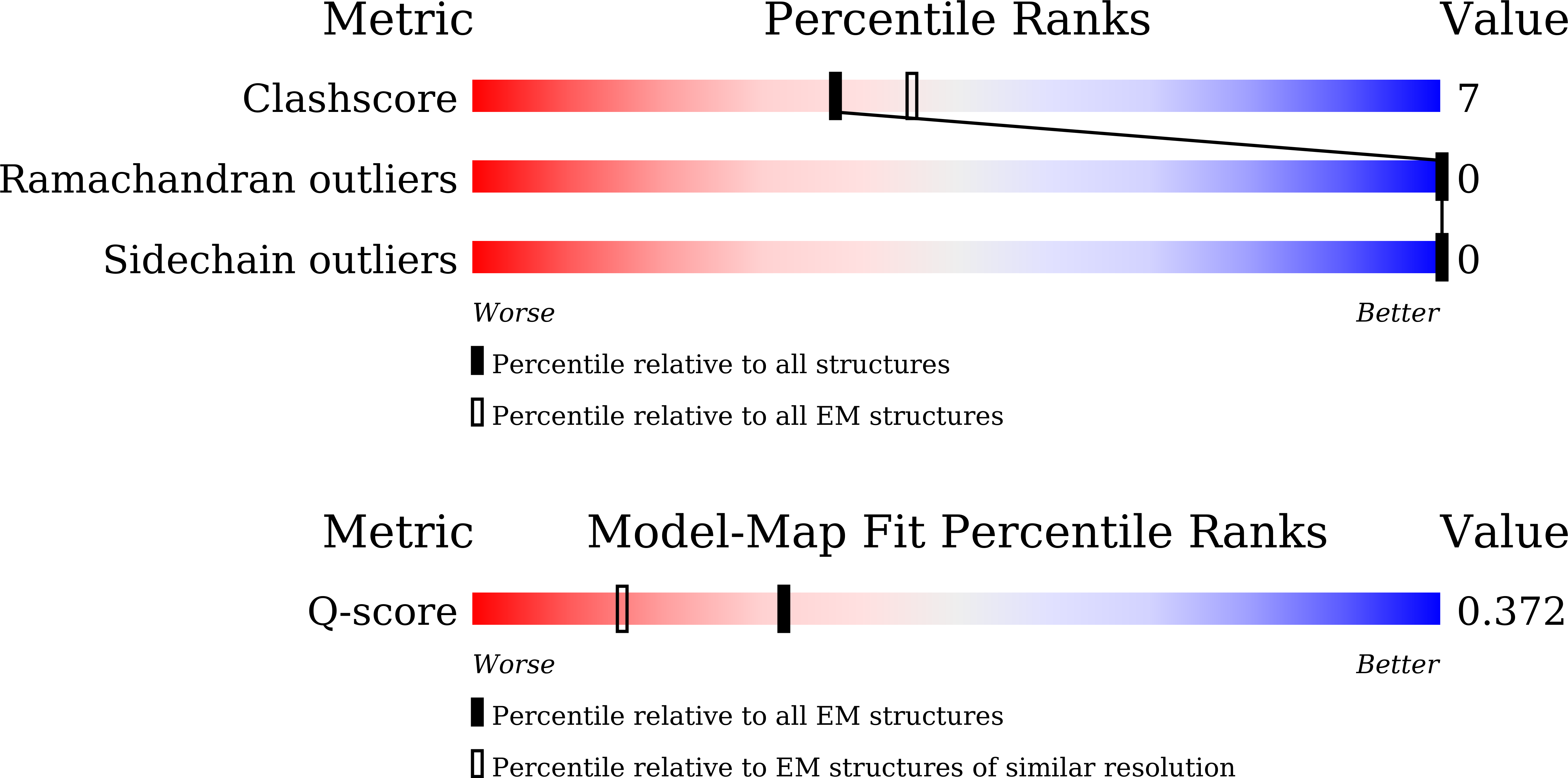
Resolution:
3.27 Å
Aggregation State:
PARTICLE
Reconstruction Method:
SINGLE PARTICLE
Macromolecular Entities
Structures with similar UniProt ID
Protein Blast
Polymer Type:polypeptide(L)
Molecule:Glutamate receptor ionotropic
Gene (Uniprot):Grin1
Chain IDs:A, C
Chain Length:821
Number of Molecules:2
Biological Source:Mus musculus
Structures with similar UniProt ID
Protein Blast
Polymer Type:polypeptide(L)
Molecule:Glutamate receptor ionotropic
Gene (Uniprot):Grin2a
Chain IDs:B
Chain Length:805
Number of Molecules:1
Biological Source:Mus musculus
Structures with similar UniProt ID
Protein Blast
Polymer Type:polypeptide(L)
Molecule:Glutamate receptor ionotropic
Gene (Uniprot):Grin2b
Chain IDs:D
Chain Length:811
Number of Molecules:1
Biological Source:Mus musculus
Primary Citation
Conformational diversity and fully opening mechanism of native NMDA receptor.
Nature 652 1405 1414 (2026)
PMID: 41673155 DOI: 10.1038/s41586-026-10139-w

Abstact

N-methyl-D-aspartate receptors (NMDARs) are glutamate-gated ion channels that mediate excitatory neurotransmission throughout the brain(1). As obligate heterotetramers, their activation requires the binding of both glycine and glutamate(2). Although recent structural studies have provided insights into endogenous receptors from select brain regions(3), most previous work has relied on recombinant receptors and engineered constructs, which limits our understanding of native NMDARs across the whole brain. Here we identify and resolve ten distinct native NMDAR assemblies from the whole-brain tissue of female C57BL/6 mice using immunoaffinity purification, single-molecule total internal reflection fluorescence microscopy and cryo-electron microscopy. Analyses of the GluN1-GluN2A(S1), GluN1-GluN2A(S2), GluN1-GluN2A(S3), GluN1-GluN2B, GluN1-GluN2A-GluN2B(S1), GluN1-GluN2A-GluN2B(S2), GluN1-GluN2A-GluNX(S1), GluN1-GluN2A-GluNX(S2), GluN1-GluN2B-GluNX and GluN1-GluNX structures reveal that GluN2A is the most prevalent subunit across assemblies. Moreover, the substantial conformational flexibility observed in the GluN2A amino-terminal domain may explain its fast kinetics and dominant role in gating. Dynamic movements of S-ketamine were also captured at the channel vestibule, as was pore dilation in both the GluN1 and GluN2B subunits of a native GluN1-GluN2B receptor. The latter observation represents a previously unknown fully open state of NMDAR. Our large collection of heterogeneous NMDAR structures from whole brain reveals previously unrecognized properties of conformational diversity and channel dilation.

Legend

Protein

Chemical

Disease

Primary Citation of related structures
Feedback Form
Name
Email
Institute
Feedback