

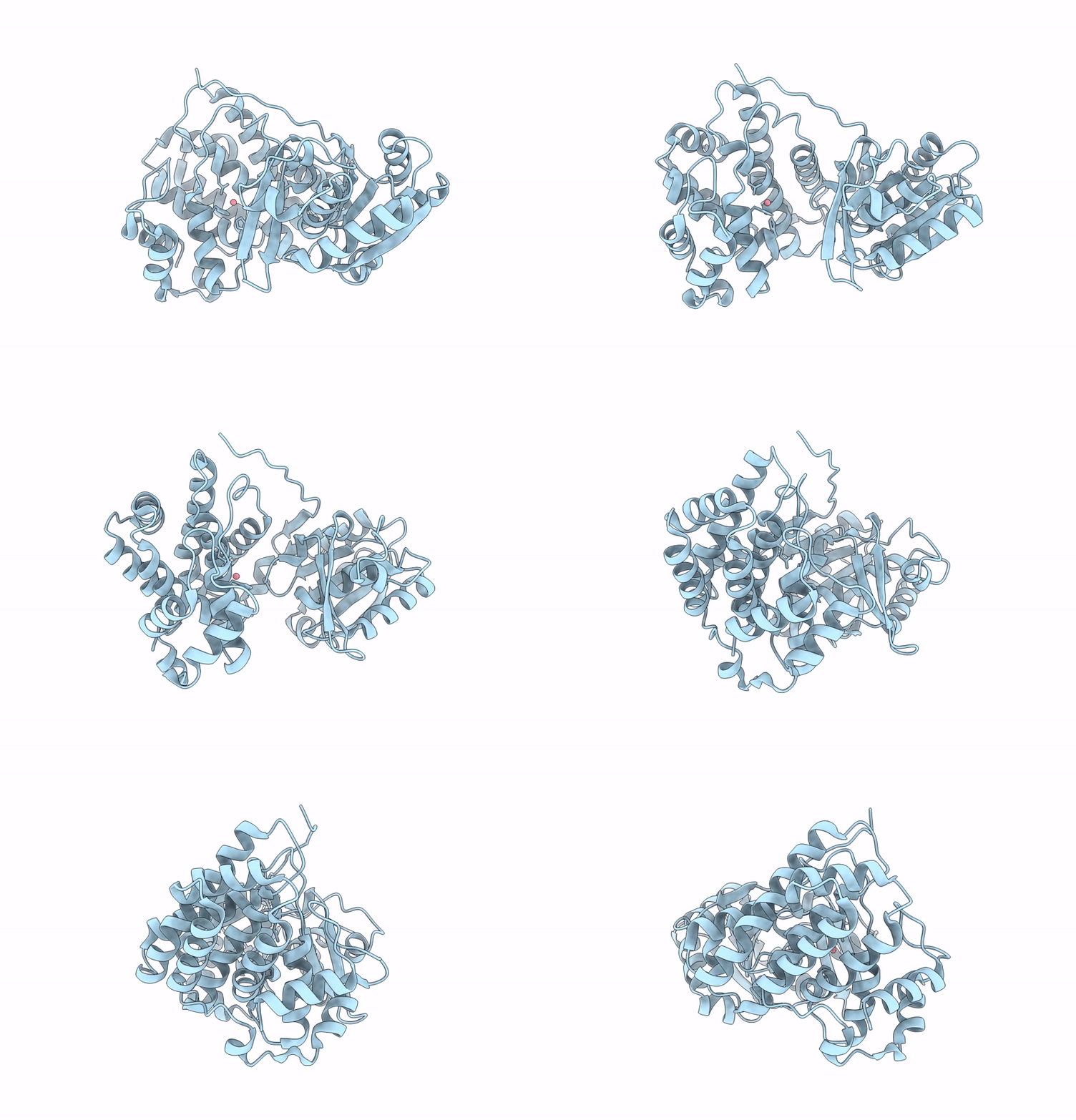
9UGS image
Deposition Date 2025-04-13
Release Date 2025-10-29
Last Version Date 2026-04-08
Entry Detail
PDB ID:
9UGS
Title:
Butanol Dehydrogenase
Biological Source:
Source Organism(s):
Expression System(s):
Method Details:
Experimental Method:
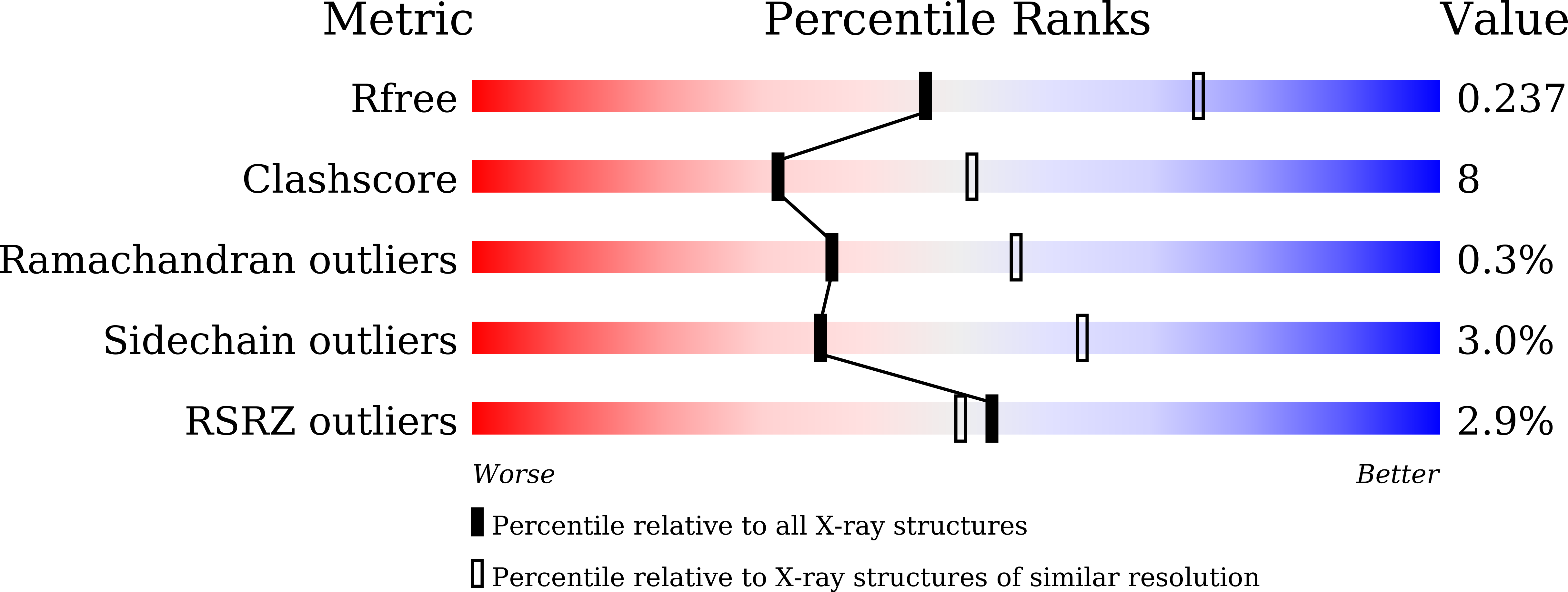
Resolution:
2.50 Å
R-Value Free:
0.23
R-Value Work:
0.18
R-Value Observed:
0.19
Space Group:
I 2 2 2
Macromolecular Entities
Structures with similar UniProt ID
Protein Blast
Polymer Type:polypeptide(L)
Molecule:NADH-dependent butanol dehydr
Gene (Uniprot):C7Y58_10070
Chain IDs:A
Chain Length:385
Number of Molecules:1
Biological Source:Fusobacterium nucleatum
Ligand Molecules
Primary Citation
Structural flexibility of the nicotinamide group of NADH in butanol dehydrogenase YqdH from Fusobacterium nucleatum.
PLoS ONE 20 e0338369 e0338369 (2025)
PMID: 41406140 DOI: 10.1371/journal.pone.0338369

Abstact

Butanol dehydrogenases (BDHs) are NAD(P)H-dependent oxidoreductases that catalyze the reversible conversion of butanol to butyraldehyde. These enzymes play essential roles in microbial butanol fermentation and show significant potential for biofuel synthesis and bioremediation. The crystal structures of BDHs from Fusobacterium nucleatum and Thermotoga maritima have clarified cofactor recognition and proposed reaction mechanisms. However, their distinct cofactor-binding modes and conformational differences in the substrate-binding cleft remain poorly characterized. In this study, we report the crystal structure of Fusobacterium nucleatum butanol dehydrogenase YqdH (FnYqdH) in a partially NADH-bound state. Electron density map analysis showed stable binding of the adenosine and diphosphate groups of NADH to the nucleotide-binding domain of FnYqdH. Conversely, the nicotinamide group was not observed, indicating that it was in an unbound state. Structural comparisons of FnYqdH complexed with either partial ADP or NADH revealed that the adenosine group is stabilized by hydrogen bonds with Thr143, Thr187, and Val184. Nicotinamide group binding induces positional and conformational changes in the diphosphate group of NADH. A comparative analysis of FnYqdH and TmBDH proteins revealed distinct conformational differences between their nucleotide-binding and catalytic domains, including variations in their substrate-binding metal ion sites. In particular, amino acid sequence and structural analyses of the BDH family revealed significant variability in the residues responsible for metal ion binding. Based on the observed flexibility of the nicotinamide group of NADH and the open conformation of FnYqdH, a potential reaction mechanism of FnYqdH is proposed. These findings offer valuable insights into the cofactor and substrate recognition within the BDH protein family.

Legend

Protein

Chemical

Disease

Primary Citation of related structures
Feedback Form
Name
Email
Institute
Feedback