Deposition Date
2025-04-10
Release Date
2026-05-13
Last Version Date
2026-05-13
Entry Detail
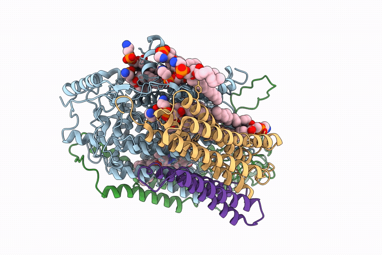
Biological Source:
Source Organism(s):
Acinetobacter baumannii (Taxon ID: 470)
Expression System(s):
Method Details:
Experimental Method:
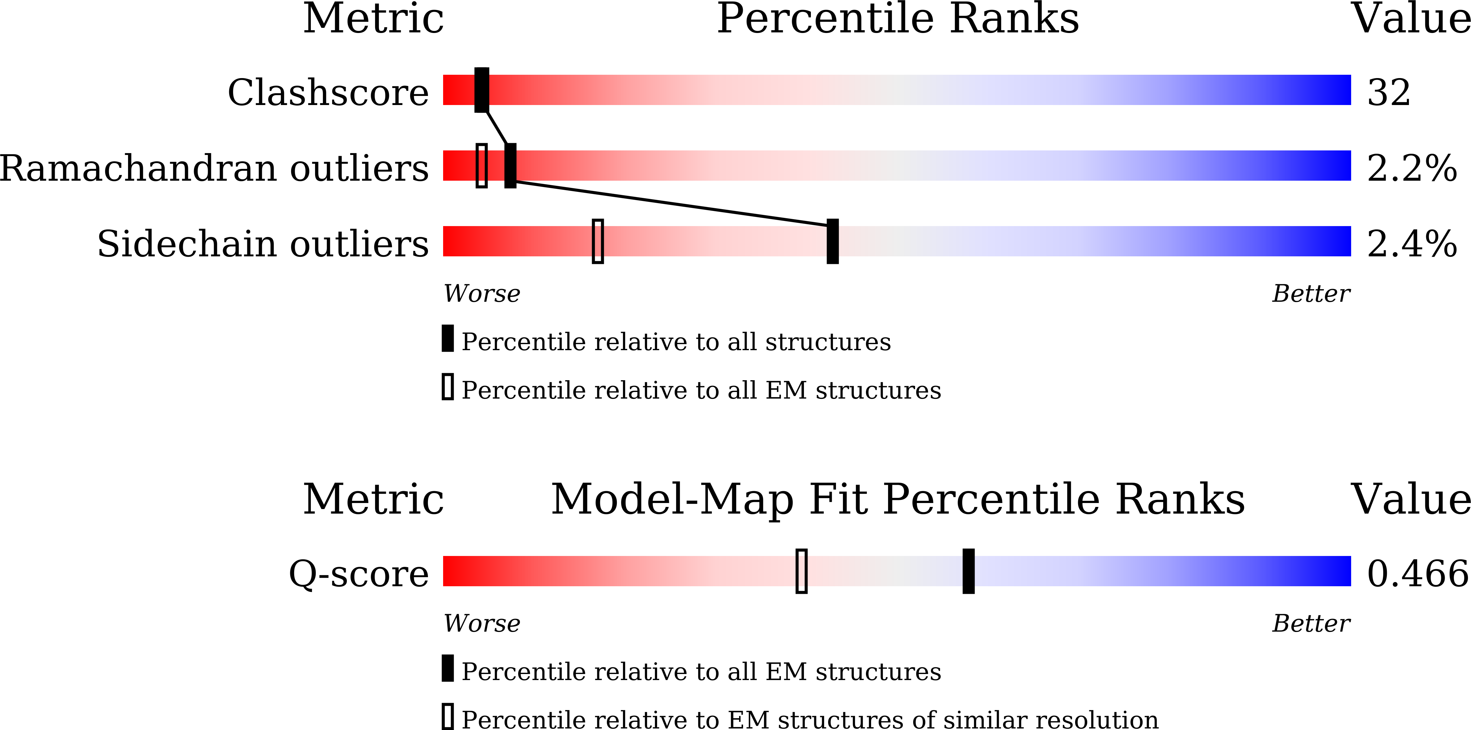
Resolution:
3.05 Å
Aggregation State:
PARTICLE
Reconstruction Method:
SINGLE PARTICLE