

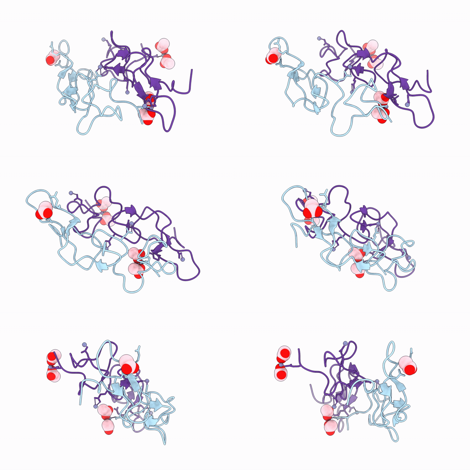
9UAL image
Deposition Date 2025-04-01
Release Date 2026-04-08
Last Version Date 2026-05-20
Entry Detail
PDB ID:
9UAL
Keywords:
Title:
Crystal structure of Trimucrin
Biological Source:
Source Organism(s):
Expression System(s):
Method Details:
Experimental Method:
Resolution:
1.68 Å
R-Value Free:
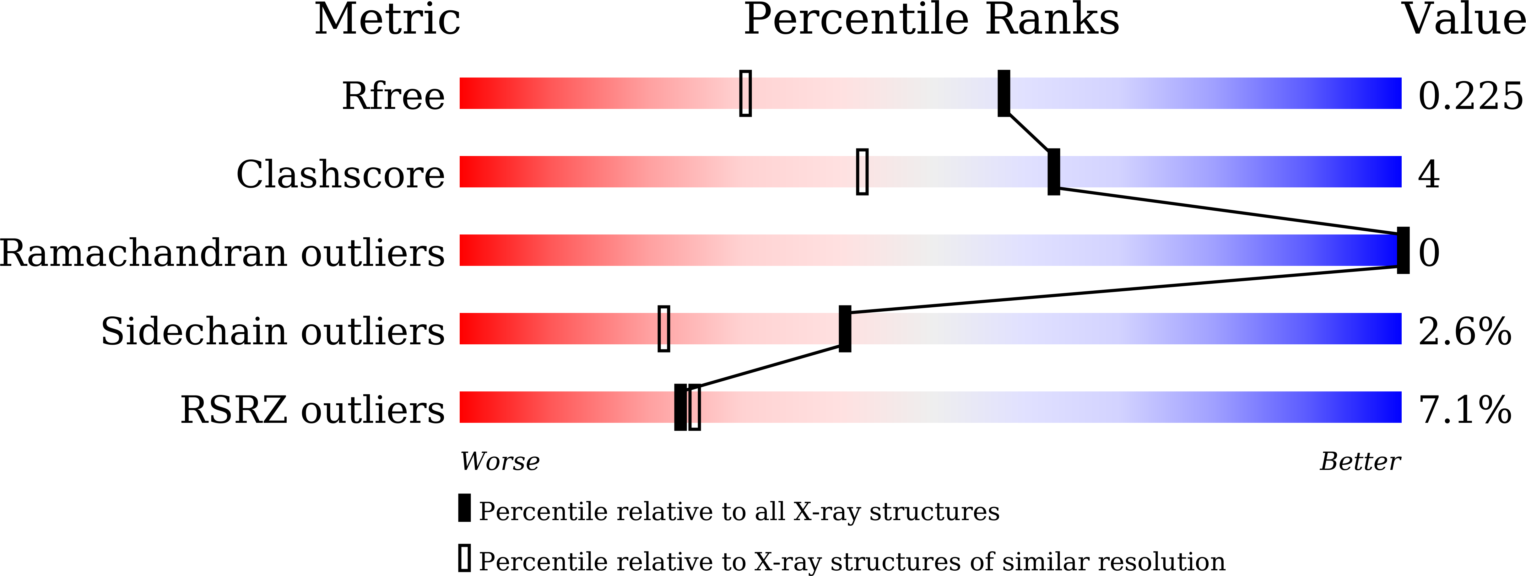
0.21
R-Value Work:
0.17
Space Group:
P 1 21 1
Macromolecular Entities
Structures with similar UniProt ID
Protein Blast
Polymer Type:polypeptide(L)
Molecule:Zinc metalloproteinase/disint
Chain IDs:A, B
Chain Length:73
Number of Molecules:2
Biological Source:Protobothrops mucrosquamatus
Primary Citation
Structural basis for the differential recognition of integrin alpha v beta 3 by rhodostomin and trimucrin.
Commun Biol 9 ? ? (2026)
PMID: 42045602 DOI: 10.1038/s42003-026-10139-6

Abstact

Rhodostomin (Rho) and Trimucrin (Tmu) are RGD-containing disintegrins that inhibit integrins more effectively than short RGD peptides. They differ in their linker, RGD loop, and C-terminal sequences. We determined the X-ray structure of Tmu and the cryo-EM structures of integrin alphavbeta3 in complex with both disintegrins. Structural analysis revealed subtle differences in binding, with both adopting a rigid backbone conformation and interacting with integrin through three cooperative binding sites. Besides the conserved RGD interface, Tmu features a cluster of basic residues in its linker, while Rho has distinct C-terminal interactions. Disintegrin binding stabilizes alphavbeta3 in an extended-open conformation, while the beta3-Y110 residue is essential for maintaining the bent state without ligands. These findings enhance our understanding of integrin recognition and inform the development of integrin-targeted therapeutics for anti-angiogenic, anti-tumor, and anti-inflammatory applications.

Legend

Protein

Chemical

Disease

Primary Citation of related structures
Feedback Form
Name
Email
Institute
Feedback