Deposition Date
2025-12-15
Release Date
2026-05-13
Last Version Date
2026-05-13
Entry Detail
PDB ID:
9TNF
Keywords:
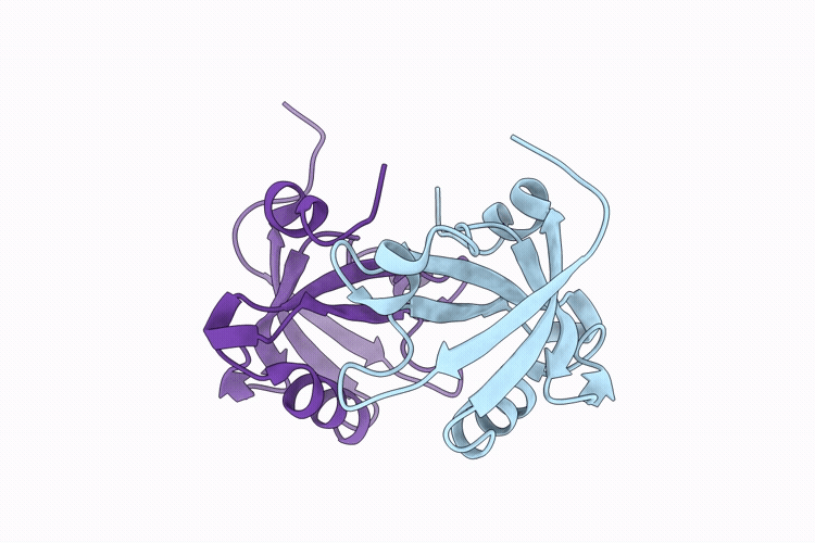
Title:
Crystal Structure of the third PDZ domain of PSD-95 protein Y397E mutant
Biological Source:
Source Organism(s):
Homo sapiens (Taxon ID: 9606)
Expression System(s):
Method Details:
Experimental Method:
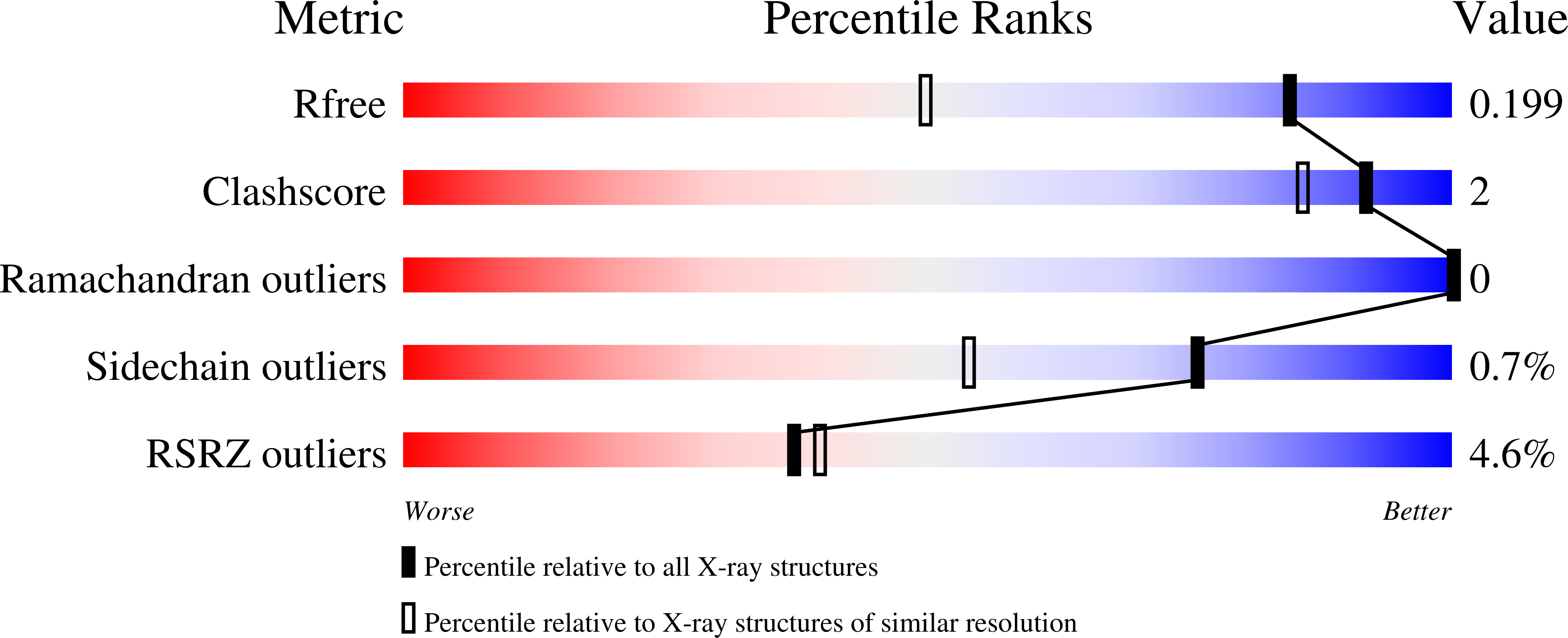
Resolution:
1.45 Å
R-Value Free:
0.20
R-Value Work:
0.16
R-Value Observed:
0.16
Space Group:
P 21 21 21