

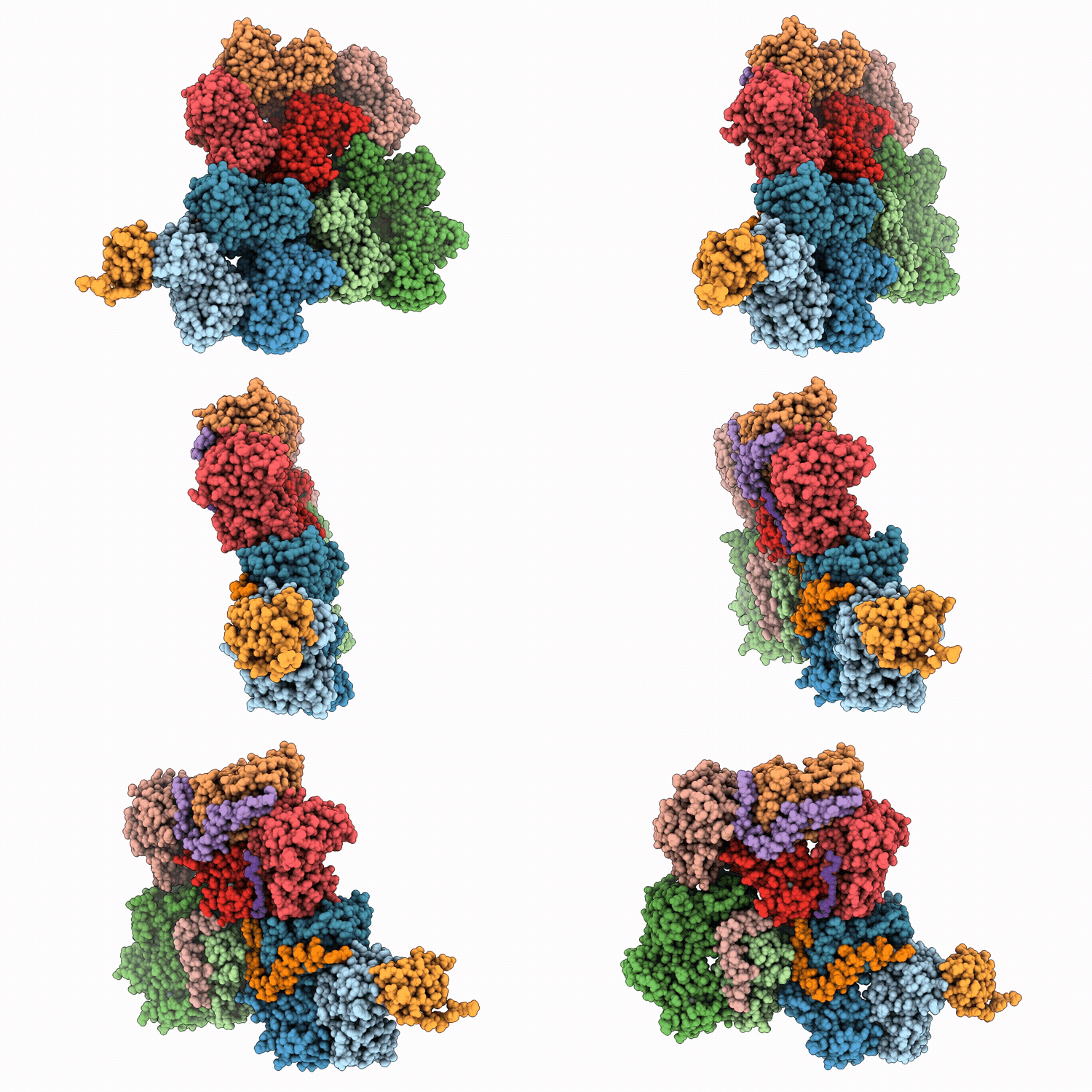
9T9V image
Deposition Date 2025-11-17
Release Date 2026-08-19
Last Version Date 2026-08-19
Entry Detail
PDB ID:
9T9V
Keywords:
Title:
Structure of bacteriophage NO16
Biological Source:
Source Organism(s):
Method Details:
Experimental Method:
Resolution:
3.30 Å
Aggregation State:
PARTICLE
Reconstruction Method:
SINGLE PARTICLE
Macromolecular Entities
Polymer Type:polypeptide(L)
Molecule:Double jelly roll capsid prot
Chain IDs:A, B, C, D, E, F, G, H, I, J
Chain Length:265
Number of Molecules:10
Biological Source:Vibrio phage fNo16
Polymer Type:polypeptide(L)
Molecule:Penton base (GP14)
Chain IDs:K
Chain Length:180
Number of Molecules:1
Biological Source:Vibrio phage fNo16
Polymer Type:polypeptide(L)
Molecule:LPXTG cell wall anchor domain
Chain IDs:L, M, N
Chain Length:82
Number of Molecules:3
Biological Source:Vibrio phage fNo16
Polymer Type:polypeptide(L)
Molecule:Unknown1
Chain IDs:O
Chain Length:12
Number of Molecules:1
Biological Source:Vibrio phage fNo16
Ligand Molecules
Primary Citation
Structure of NO16, a marine non-tailed vibriophage with an unusual symmetry-mismatched vertex arrangement
To Be Published ? ? ? (2026)
DOI: 10.1371/journal.ppat.1014525
Primary Citation of related structures
Feedback Form
Name
Email
Institute
Feedback