9SOJ image
Deposition Date 2025-09-13
Release Date 2026-04-22
Last Version Date 2026-05-06
Entry Detail
PDB ID:
9SOJ
Title:
Protein with novel 3_1 knotted solenoid fold
Biological Source:
Source Organism(s):
Expression System(s):
Method Details:
Experimental Method:
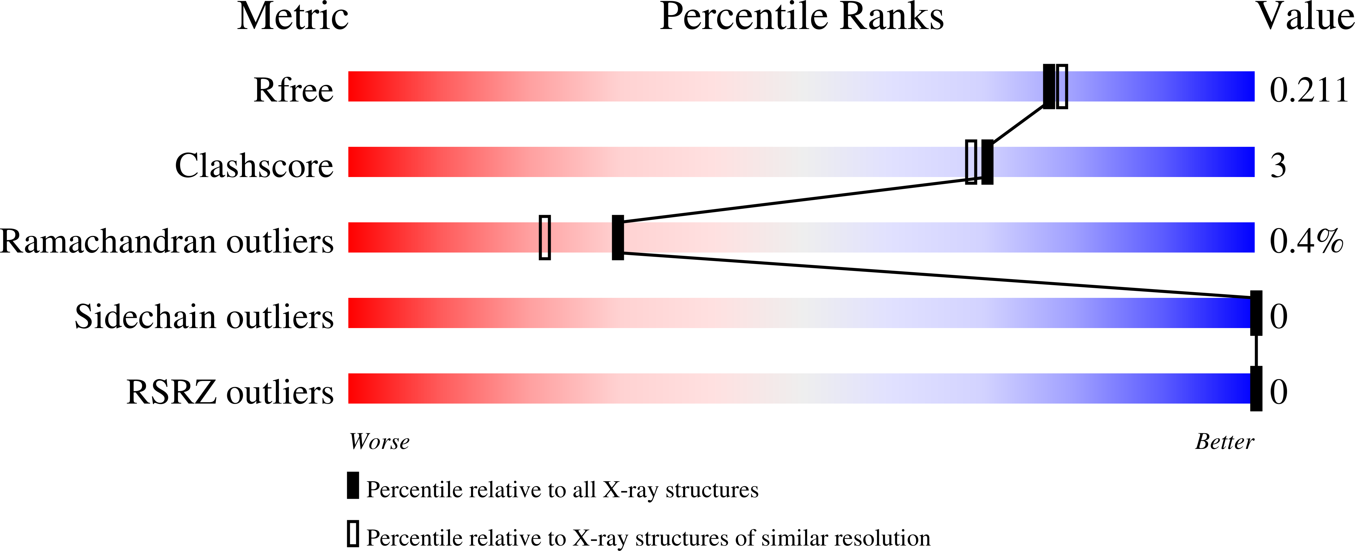
Resolution:
1.90 Å
R-Value Free:
0.21
R-Value Work:
0.16
R-Value Observed:
0.16
Space Group:
P 21 21 2
Macromolecular Entities
Structures with similar UniProt ID
Protein Blast
Polymer Type:polypeptide(L)
Molecule:Novel 3_1 knotted solenoid
Chain IDs:A, B, C, D
Chain Length:139
Number of Molecules:4
Biological Source:Brevundimonas sp. SH203
Ligand Molecules
Primary Citation
Novel Knotted Solenoid fold with order-shifted coil arrangement leads to nontrivial 3 1 topology.
Proc.Natl.Acad.Sci.USA 123 e2525920123 e2525920123 (2026)
PMID: 42018416 DOI: 10.1073/pnas.2525920123

Abstact

Herein, we present crystal structures of proteins adopting a fold not identified among known [Formula: see text]-solenoids in current structural databases, a Knotted Solenoid. These proteins exhibit a characteristic solenoidal architecture, closely resembling [Formula: see text]-solenoid proteins. However, a unique "skip-and-backtrack" shift is observed: One coil skips a rotation while the next realigns, distinguishing this fold from previously described solenoids and allowing the formation of a 3(1) (trefoil) knot. Moreover, the proteins form homodimers, and conservation of interface residues suggests a shared oligomerization state across all Knotted Solenoids. This fold is exclusive to a specific group of bacteria and remains structurally conserved despite high sequence variability (pairwise identities down to 6%). Conserved residues are observed at the beginning of each coil and within the knot core, suggesting functional or structural significance, and an independent evolutionary path to unknotted solenoids. In vitro chemical and thermal stability studies showed fully reversible unfolding in urea, and no additional transitions even in high concentrations of guanidinium chloride. The far-ultraviolet circular dichroism unfolding kinetics showed relatively rapid unfolding. Explicit-solvent molecular dynamics simulations and a generative deep learning model show that topological constraints stabilize "skip-and-backtrack" shift in the Knotted Solenoid. A monomeric unit can self-tie into the native 3(1) knotted state through a slipknot intermediate. It then interacts with another chain via its hydrophobic surface, promoting second chain folding and dimerization. The identification of novel knotted proteins within a previously considered unknotted fold provides an opportunity to investigate the evolutionary pressures and functional implications of knotting in shaping protein architecture.

Legend

Protein

Chemical

Disease

Primary Citation of related structures
Feedback Form
Name
Email
Institute
Feedback