

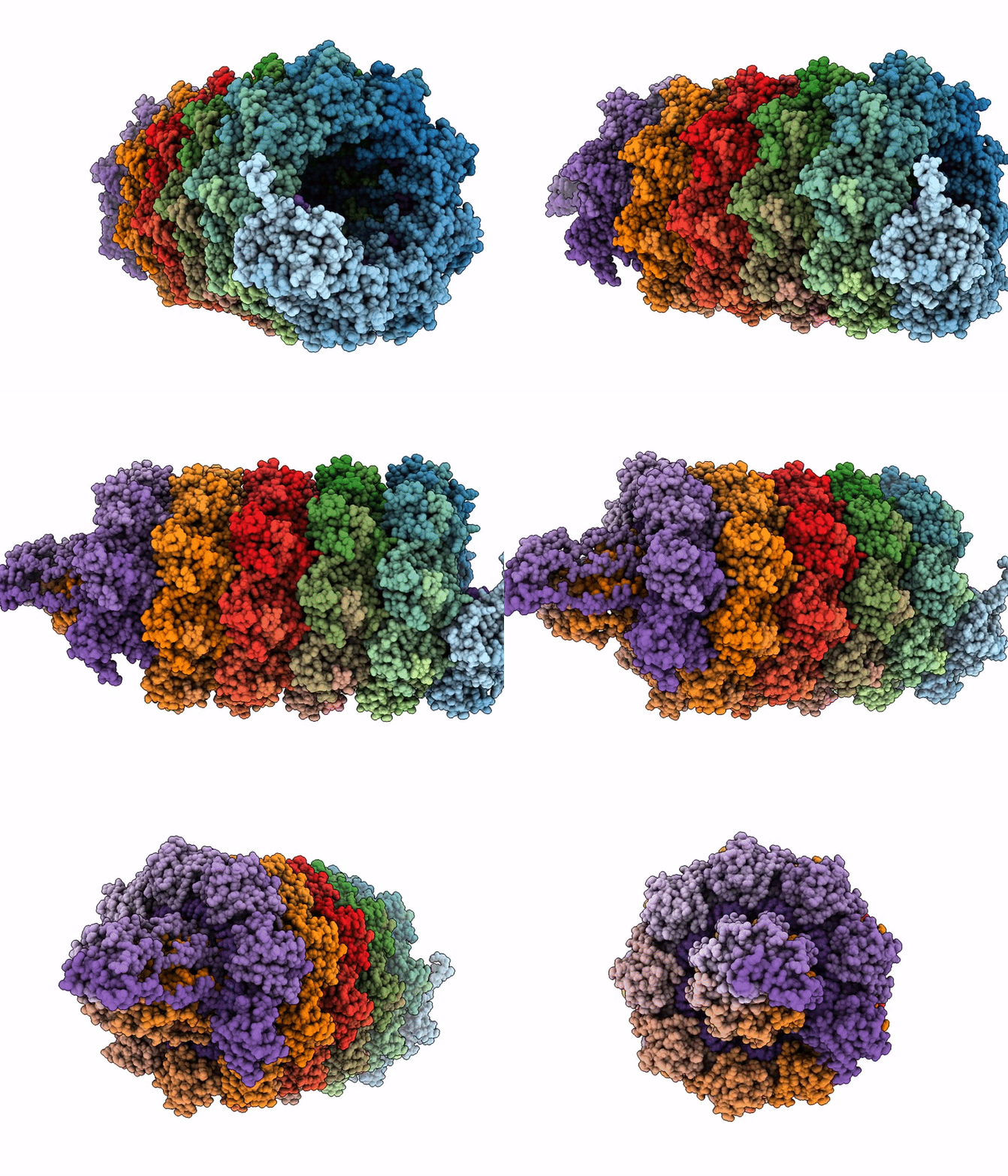
9QY3 image
Deposition Date 2025-04-16
Release Date 2026-02-25
Last Version Date 2026-02-25
Entry Detail
PDB ID:
9QY3
Keywords:
Title:
Structure of the Plum Pox Virus (PPV)
Biological Source:
Source Organism(s):
Plum pox virus (Taxon ID: 12211)
Expression System(s):
Method Details:
Experimental Method:
Resolution:
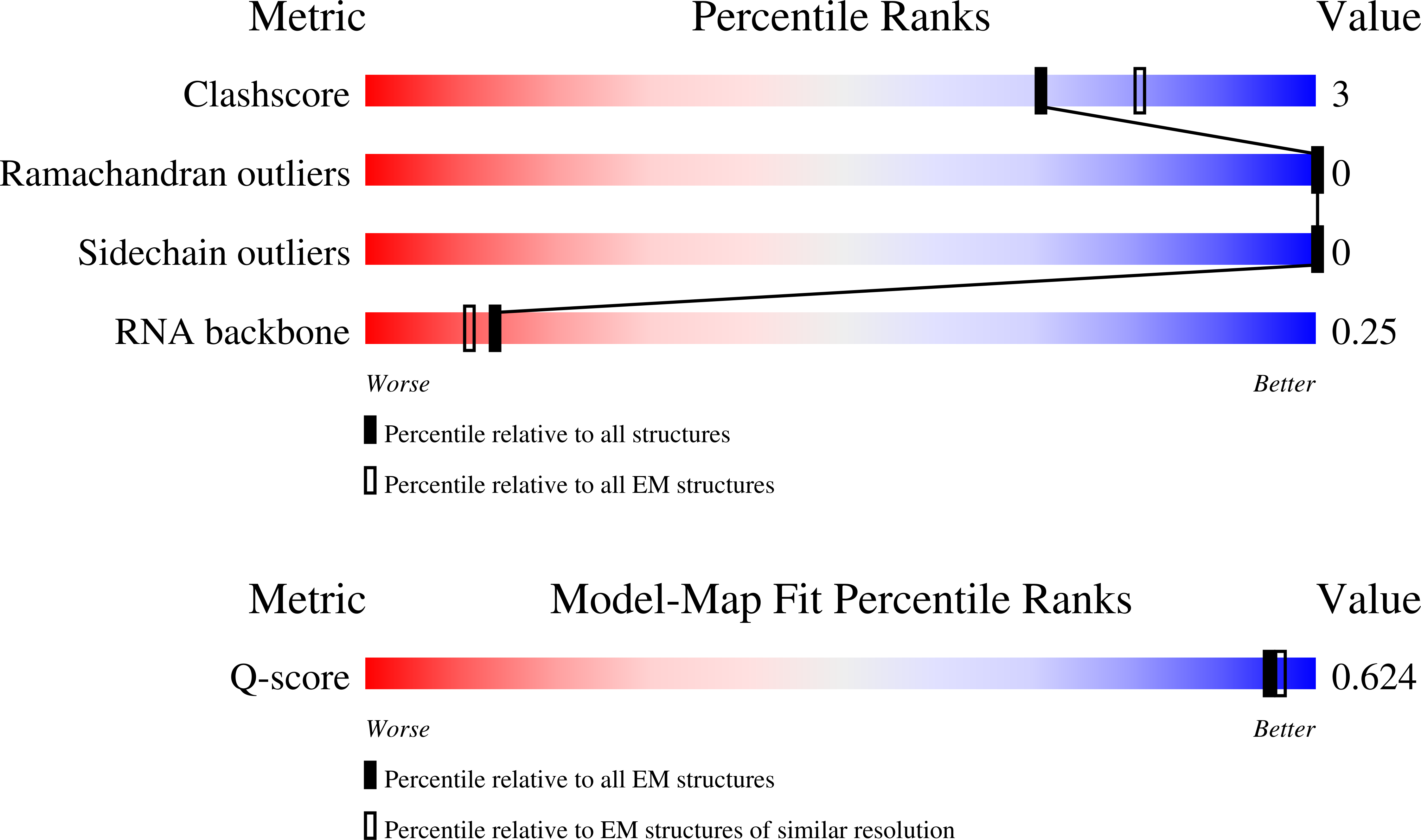
2.90 Å
Aggregation State:
FILAMENT
Reconstruction Method:
HELICAL
Macromolecular Entities
Polymer Type:polypeptide(L)
Molecule:Coat Protein (CP)
Chain IDs:A, B, C, D, E, F, G, H, I, J, K, L, M, N, O, P, Q, R, S, T, U, V, W, X, Y, Z, AA (auth: a), BA (auth: b), CA (auth: c), DA (auth: d), EA (auth: e), FA (auth: f), GA (auth: g), HA (auth: h), IA (auth: i), JA (auth: j), KA (auth: k), LA (auth: l), MA (auth: m), NA (auth: n), OA (auth: o), PA (auth: p), QA (auth: q), RA (auth: r), SA (auth: s)
Chain Length:330
Number of Molecules:45
Biological Source:Plum pox virus
Polymer Type:polyribonucleotide
Molecule:RNA
Chain IDs:TA (auth: t)
Chain Length:624
Number of Molecules:1
Biological Source:Plum pox virus
Ligand Molecules
Primary Citation
Structural characterization of plum pox virus by cryo-electron microscopy.
Arch Virol 171 11 11 (2025)
PMID: 41326719 DOI: 10.1007/s00705-025-06473-5

Abstact

Plum pox virus (PPV), a significant member of the genus Potyvirus, represents a global agricultural challenge, causing significant economic losses and threatening fruit farming due to its easy transmission to most Prunus species. Here, we present the high-resolution structural characterization of PPV using cryo-electron microscopy (cryo-EM). The reconstructed structure at 2.9 A reveals a filamentous virion with a helical assembly formed by the coat protein (CP), which encapsidates a single-stranded RNA (ssRNA) genome. The structure of the CP core shows remarkable conservation with other potyviruses, with an RNA binding site and inter-subunit interactions mediated in part by the N-terminal arm, which is confirmed here to have a disordered structure. Mass spectrometry analysis identified numerous post-translational modifications, mostly phosphorylation, primarily in the flexible N-terminal region. In silico predictions revealed intrinsically disordered regions, which is compatible with the amyloidogenic properties of the CP. These results provide new insights into the architecture and assembly of PPV, offering a basis for future studies and, possibly, antiviral strategies.

Legend

Protein

Chemical

Disease

Primary Citation of related structures
Feedback Form
Name
Email
Institute
Feedback