

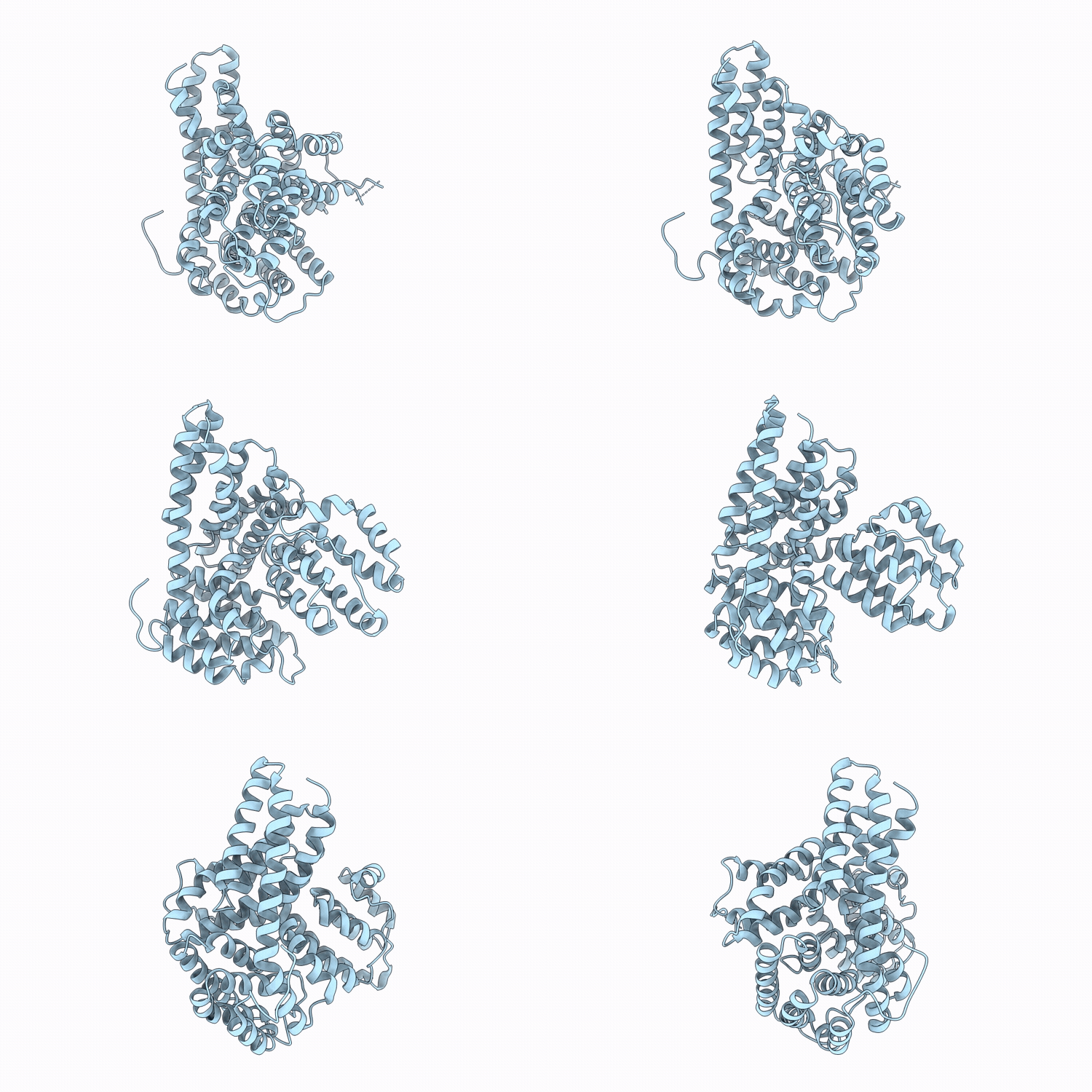
9QVI image
Deposition Date 2025-04-11
Release Date 2026-04-22
Last Version Date 2026-09-30
Entry Detail
PDB ID:
9QVI
Title:
Central domain of Glucan Water Dikinase-1 in alternative closed conformation
Biological Source:
Source Organism(s):
Expression System(s):
Method Details:
Experimental Method:
Resolution:
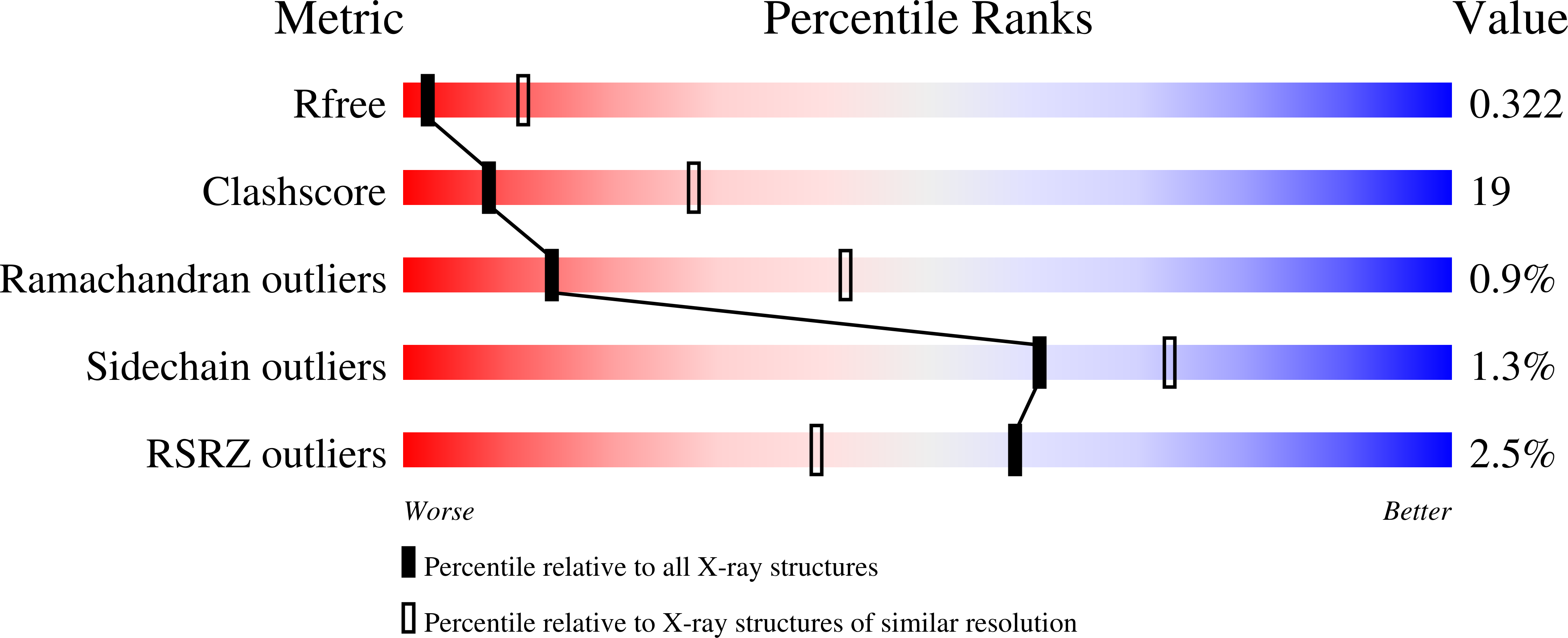
3.25 Å
R-Value Free:
0.30
R-Value Work:
0.26
Space Group:
P 43 21 2
Macromolecular Entities
Polymer Type:polypeptide(L)
Molecule:Alpha-glucan water dikinase,
Gene (Uniprot):R1
Chain IDs:A
Chain Length:465
Number of Molecules:1
Biological Source:Solanum tuberosum
Ligand Molecules
Primary Citation
A pivoting histidine domain mediates phosphate transfer in glucan water dikinase.
J.Biol.Chem. 302 113416 113416 (2026)
PMID: 42567505 DOI: 10.1016/j.jbc.2026.113416

Abstact

As the sole naturally occurring covalent modification of starch, phosphorylation plays a critical role in regulating starch metabolism across higher plants and algae. Starch phosphorylation is catalyzed by high molar mass dikinases, such as the glucan water dikinase 1 from Solanum tuberosum (StGWD1). This is the most extensively studied glucan dikinase, and preliminary structure prediction and comparison with other dikinases suggested a swiveling mechanism for beta-phosphate transfer to the glucan substrate; however, the experimental 3D structure of StGWD1 remains largely unknown, and its structural dynamics lack experimental validation. Here, we employed biochemical characterization, AlphaFold2 modeling, X-ray crystallography, and Small-Angle X-ray Scattering to gain insight into the structure and mechanism of StGWD1. The protein comprises five domains, including two N-terminal carbohydrate binding domains followed by a central domain, whose structure was solved by X-ray crystallography in both open and closed conformations. They are followed by the domain bearing the catalytic histidine and the ATP-binding domain. Using Small-Angle X-ray Scattering -driven modeling, we characterized the spatial arrangement of the full-length enzyme and several truncated variants, identifying a pivoting movement of the histidine domain consistent with autophosphorylation and subsequent phosphate transfer to glucan. Our data highlight residues at the domain interfaces that may assist catalysis. Furthermore, we hypothesize that the second carbohydrate binding domain, which remains always close to the central domain, helps maintaining the catalytic domain in proximity to the glucan chain for productive phosphate transfer.

Legend

Protein

Chemical

Disease

Primary Citation of related structures
Feedback Form
Name
Email
Institute
Feedback