Deposition Date
2025-07-18
Release Date
2026-06-10
Last Version Date
2026-06-10
Entry Detail
PDB ID:
9PN1
Keywords:
Title:
Crystal structure of Q108K:K40L:T51V:T53C:R58W:T29L:Y19W:Q4A mutant of cellular retinol binding protein II complex with 15-cis-retinal
Biological Source:
Source Organism(s):
Homo sapiens (Taxon ID: 9606)
Expression System(s):
Method Details:
Experimental Method:
Resolution:
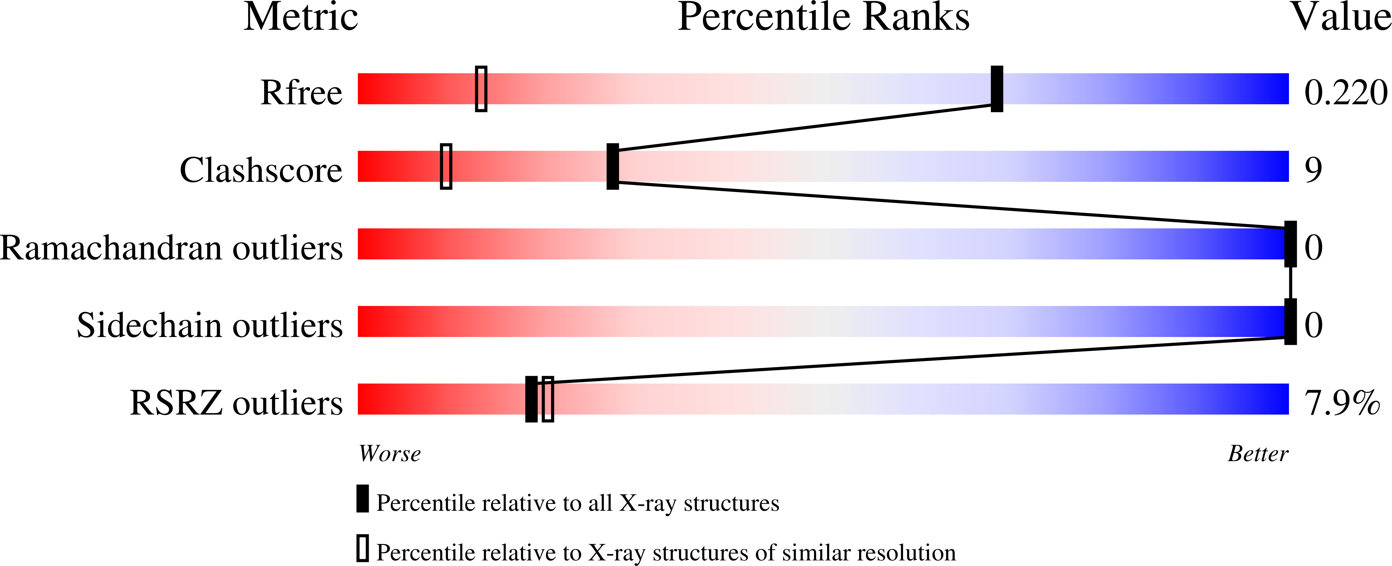
1.26 Å
R-Value Free:
0.22
R-Value Work:
0.19
R-Value Observed:
0.19
Space Group:
P 1