9PGH image
Deposition Date 2025-07-07
Release Date 2026-02-25
Last Version Date 2026-04-15
Entry Detail
PDB ID:
9PGH
Title:
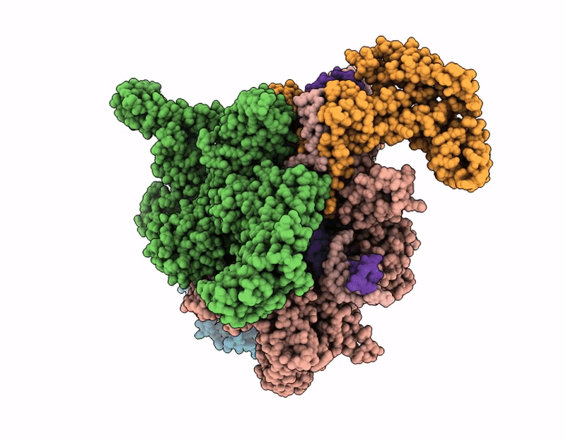
The cryo-EM structure of C. crescentus RNAP-Sigma73-CCNA_03891/CCNA_01149 promoter complex
Biological Source:
Source Organism(s):
Expression System(s):
Method Details:
Experimental Method:
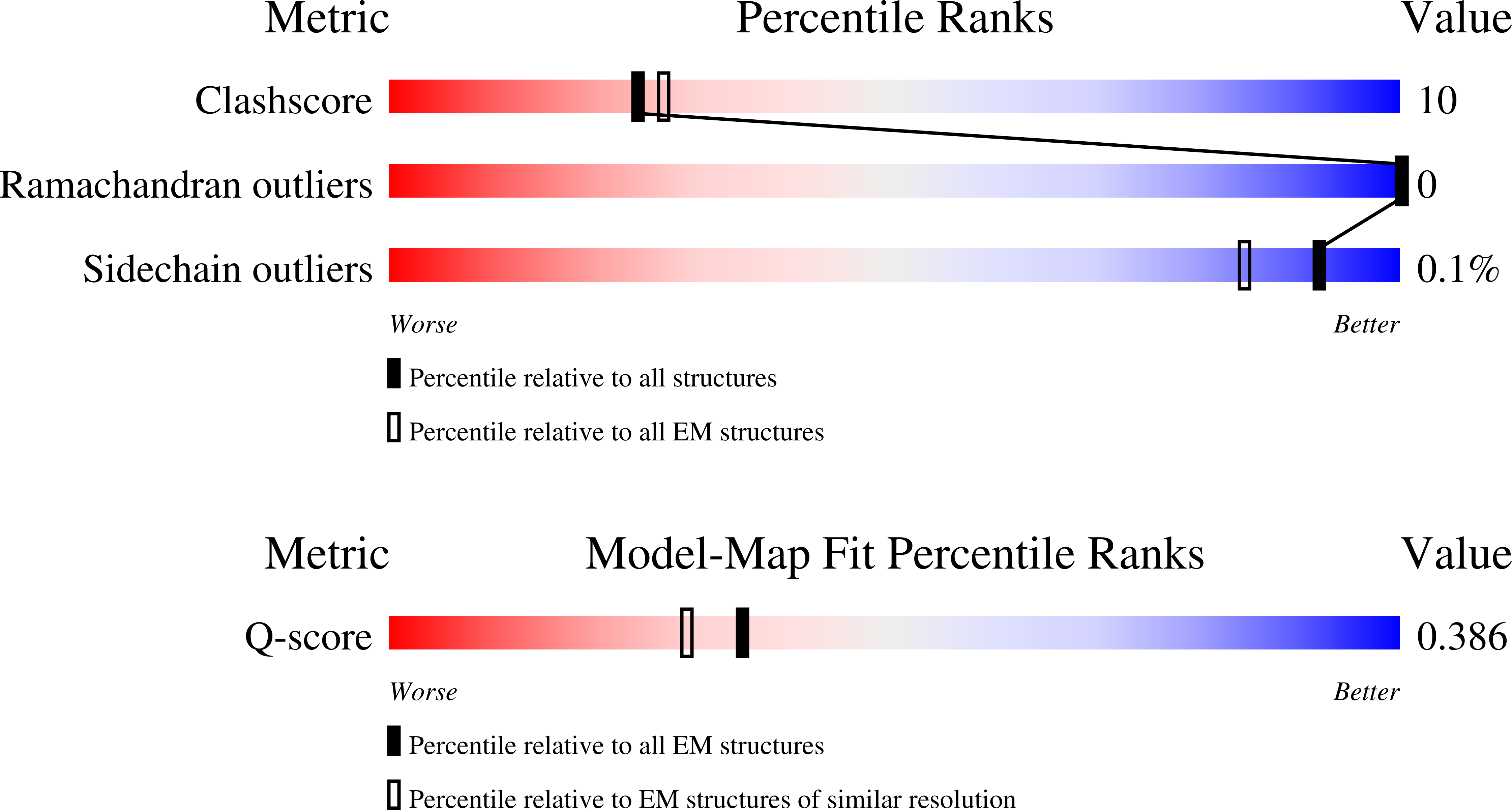
Resolution:
3.54 Å
Aggregation State:
PARTICLE
Reconstruction Method:
SINGLE PARTICLE
Macromolecular Entities
Structures with similar UniProt ID
Protein Blast
Polymer Type:polypeptide(L)
Molecule:DNA-directed RNA polymerase s
Gene (Uniprot):rpoA
Chain IDs:A, B
Chain Length:338
Number of Molecules:2
Biological Source:Caulobacter vibrioides NA1000
Structures with similar UniProt ID
Protein Blast
Polymer Type:polypeptide(L)
Molecule:DNA-directed RNA polymerase s
Gene (Uniprot):rpoB
Chain IDs:C
Chain Length:1356
Number of Molecules:1
Biological Source:Caulobacter vibrioides NA1000
Structures with similar UniProt ID
Protein Blast
Polymer Type:polypeptide(L)
Molecule:DNA-directed RNA polymerase s
Gene (Uniprot):rpoC
Chain IDs:D
Chain Length:1396
Number of Molecules:1
Biological Source:Caulobacter vibrioides NA1000
Structures with similar UniProt ID
Protein Blast
Polymer Type:polypeptide(L)
Molecule:DNA-directed RNA polymerase s
Gene (Uniprot):rpoZ
Chain IDs:E
Chain Length:119
Number of Molecules:1
Biological Source:Caulobacter vibrioides NA1000
Structures with similar UniProt ID
Protein Blast
Polymer Type:polypeptide(L)
Molecule:RNA polymerase sigma factor R
Gene (Uniprot):rpoD
Chain IDs:F
Chain Length:652
Number of Molecules:1
Biological Source:Caulobacter vibrioides NA1000
Polymer Type:polydeoxyribonucleotide
Molecule:DNA (55-MER)-non template
Chain IDs:G (auth: H)
Chain Length:99
Number of Molecules:1
Biological Source:Caulobacter vibrioides NA1000
Polymer Type:polydeoxyribonucleotide
Molecule:DNA (43-MER)-template
Chain IDs:H (auth: I)
Chain Length:99
Number of Molecules:1
Biological Source:Caulobacter vibrioides NA1000
Primary Citation
Transcription activation mechanism of a noncanonical DNA damage response pathway by the WYL-activator, DriD.
Sci Adv 12 eaec6337 eaec6337 (2026)
PMID: 41861017 DOI: 10.1126/sciadv.aec6337

Abstact

DNA damage repair mechanisms are vital for cell survival. In the bacterium, Caulobacter crescentus, DriD is the master regulator of a unique, noncanonical DNA damage pathway. DriD binding to ssDNA, produced upon DNA damage, stimulates its ability to activate transcription from several promoters involved in DNA damage responses. However, the mechanism by which DriD interfaces with the RNAP holoenzyme to activate transcription from its multiple promoters has been unclear. Here, we describe cryo-EM structures of DriD-ssDNA bound to RNAP-holoenzyme and three DriD-regulated promoters. Each subunit of homodimeric DriD contains an DNA binding N-terminal winged helix-turn-helix (wHTH) connected to WYL domains by a linker 3-helix bundle (3HB) module. The structures reveal a mechanism of assembly on promoters whereby DriD's 3HBs bind the RNAP alpha-CTD and beta domains, anchoring the RNAP-holoenzyme to regulated promoters. The 3HBs form autoinhibitory contacts with DNABDs in apo DriD and therefore acts as an ssDNA-driven trigger domain, switching between DNABD-bound apo and RNAP-bound forms upon ssDNA-mediated activation. Thus, the structures reveal a unique transcription activation mechanism, likely conserved among the large family of homodimeric WYL activators.

Legend

Protein

Chemical

Disease

Primary Citation of related structures
Feedback Form
Name
Email
Institute
Feedback