Deposition Date
2025-06-05
Release Date
2025-12-03
Last Version Date
2026-06-24
Entry Detail
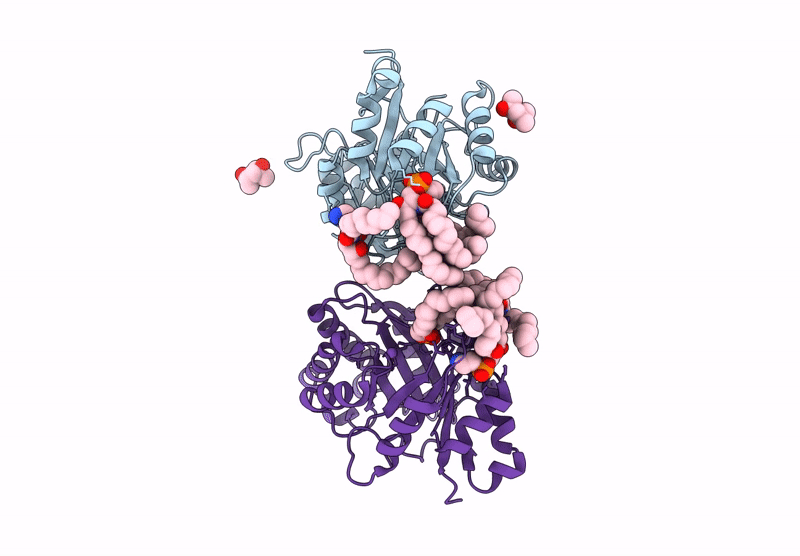
PDB ID:
9OZO
Keywords:
Title:
Structure of phospholipase D BetaIB1i from Sicarius terrosus venom, H47N mutant bound to product and substrate sphingolipids at 2.2 A resolution from a 2-day old crystal
Biological Source:
Source Organism(s):
Sicarius terrosus (Taxon ID: 571544)
Expression System(s):
Method Details:
Experimental Method:
Resolution:
2.20 Å
R-Value Free:
0.19
R-Value Work:
0.15
R-Value Observed:
0.15
Space Group:
I 1 2 1