

9O5F image
Deposition Date 2025-04-10
Release Date 2026-01-14
Last Version Date 2026-04-15
Entry Detail
PDB ID:
9O5F
Title:
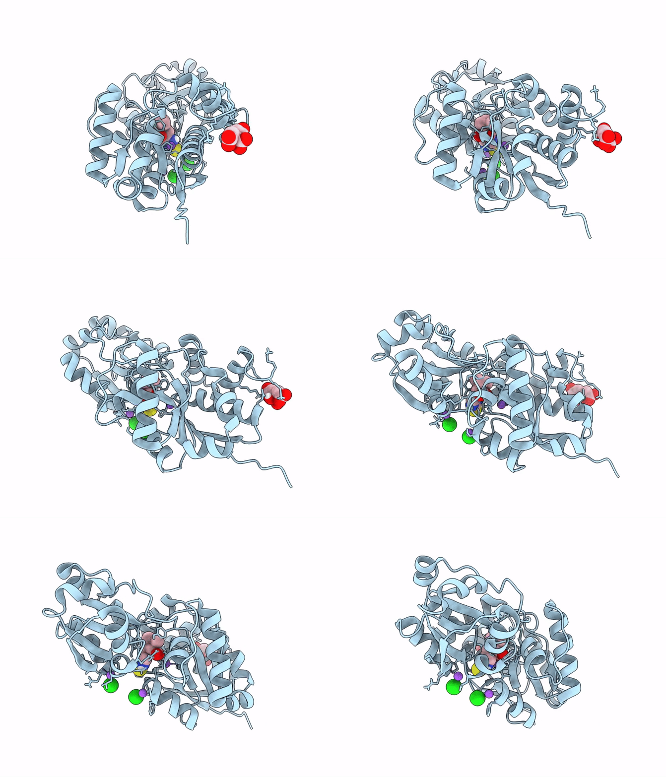
Crystal Structure of EgtUC binding domain double mutant I243P T274G bound to L-Ergothioneine from S. pneumoniae
Biological Source:
Source Organism(s):
Expression System(s):
Method Details:
Experimental Method:
Resolution:
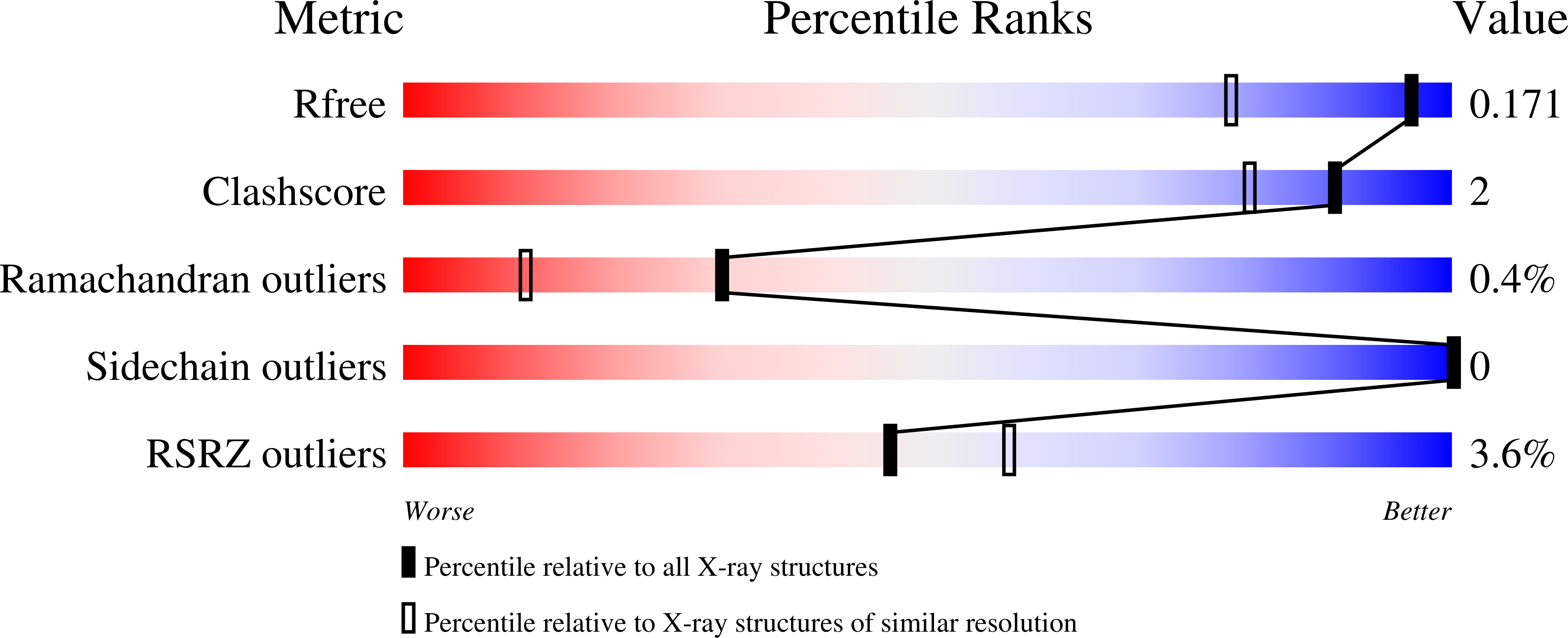
1.36 Å
R-Value Free:
0.16
R-Value Work:
0.14
R-Value Observed:
0.14
Space Group:
I 41 2 2
Macromolecular Entities
Structures with similar UniProt ID
Protein Blast
Polymer Type:polypeptide(L)
Molecule:Ergothioneine transporter Egt
Gene (Uniprot):egtUBC
Chain IDs:A
Chain Length:277
Number of Molecules:1
Biological Source:Streptococcus pneumoniae D39
Primary Citation
CH•••S hydrogen bonds drive molecular recognition of ergothioneine by the microbial transporter.
Sci Adv 12 eaeb0426 eaeb0426 (2026)
PMID: 41719410 DOI: 10.1126/sciadv.aeb0426

Abstact

Many bacteria harbor an ATP-binding cassette (ABC) transporter named EgtU specific for the human dietary antioxidant and 2-thioimidazole-containing low-molecular weight thiol ergothioneine (ET). How the solute binding domain, EgtUC, discriminates among ET and other similar molecules is unknown. Here, we use a "chimeric" mutagenesis strategy and two distantly related EgtUCs from Streptococcus pneumoniae and Helicobacter pylori to show that a suite of EgtUC alkyl CH***S hydrogen bonds to the ET thione S atom are central determinants of molecular recognition. Small perturbations in CH***S distance and angle give rise to sharply attenuated transport-competent ET-bound "closed" state lifetimes and increased motional disorder in the binding pocket, not around the S atom itself, but distally in weakening NH***O hydrogen bonds. This work highlights the impact of alkyl CH***S H bonding in a biological protein-ligand complex in water.

Legend

Protein

Chemical

Disease

Primary Citation of related structures
Feedback Form
Name
Email
Institute
Feedback