

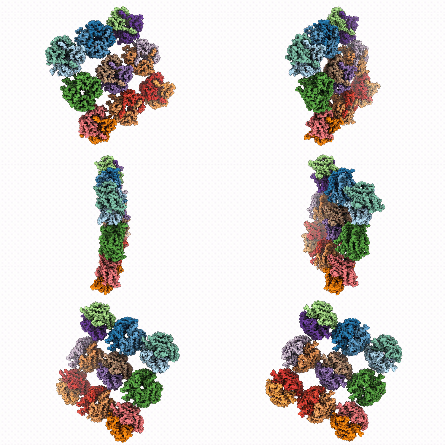
9NQY image
Deposition Date 2025-03-13
Release Date 2026-08-19
Last Version Date 2026-08-19
Entry Detail
PDB ID:
9NQY
Title:
Nipah Virus Matrix Lattice in Complex with Human Cell Membrane
Biological Source:
Source Organism(s):
Expression System(s):
Method Details:
Experimental Method:
Resolution:
7.00 Å
Aggregation State:
3D ARRAY
Reconstruction Method:
SUBTOMOGRAM AVERAGING
Macromolecular Entities
Polymer Type:polypeptide(L)
Molecule:Matrix protein
Gene (Uniprot):M
Chain IDs:A (auth: D), B (auth: A), C (auth: B), D (auth: C), E, F, G, H, I, J (auth: M), K (auth: J), L (auth: K), M (auth: L), N, O, P, Q, R
Chain Length:0
Number of Molecules:18
Biological Source:Henipavirus nipahense
Ligand Molecules
Primary Citation
Cryo-electron tomography of Nipah virus structural protein complexes in virus-like particles.
Biorxiv ? ? ? (2026)
PMID: 42523209 DOI: 10.64898/2026.07.16.738223

Abstact

Nipah virus (NiV) is a BSL-4 zoonotic paramyxovirus with ~75% human mortality. The matrix protein (M) of NiV and other paramyxoviruses binds the inner leaflet of the cellular plasma membrane, orchestrating virion assembly by bringing together transmembrane glycoproteins (F/G) and ribonucleoprotein complexes (N). However, the interactions of these full-length proteins within membrane complexes remain elusive. Using cryo-electron tomography and subtomogram averaging of virus like particles (VLPs), we interrogated the protein:protein interactions of the main NiV structural proteins M/N/F/G. The M lattice structure determined to 7A revealed a novel M-dimer arrangement that yielded two distinct repeating holes. Notably, F-trimers were arranged above only one of the two holes, dependent on F's cytoplasmic tail. G was enriched in regions of higher M-VLP curvature, while N dramatically increased M-VLP pleomorphism. This work provides novel insights into paramyxoviral protein complexes, structures, and morphology.

Legend

Protein

Chemical

Disease

Primary Citation of related structures
Feedback Form
Name
Email
Institute
Feedback