Deposition Date
2025-03-03
Release Date
2026-02-18
Last Version Date
2026-05-06
Entry Detail
PDB ID:
9NLN
Keywords:
Title:
Crystal Structure of Coxsackievirus B3 IRES Domain V in Complex with a Fab
Biological Source:
Source Organism(s):
Homo sapiens (Taxon ID: 9606)
Coxsackievirus B3 (strain Nancy) (Taxon ID: 103903)
Coxsackievirus B3 (strain Nancy) (Taxon ID: 103903)
Expression System(s):
Method Details:
Experimental Method:
Resolution:
3.00 Å
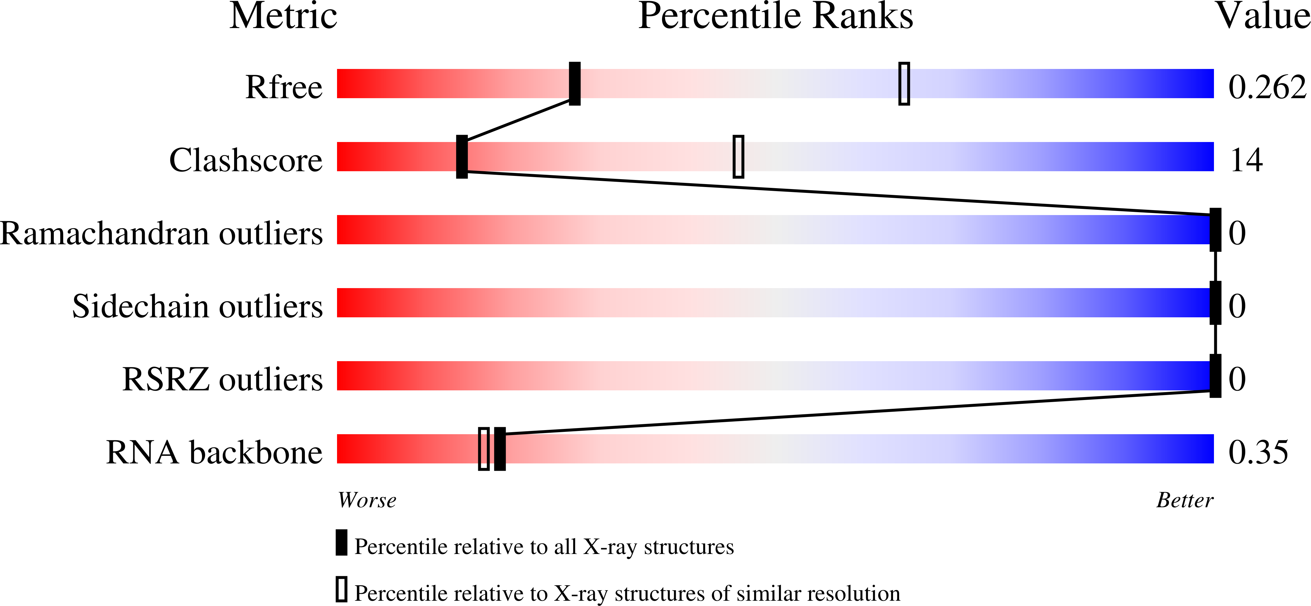
R-Value Free:
0.26
R-Value Work:
0.21
R-Value Observed:
0.21
Space Group:
P 1 21 1