

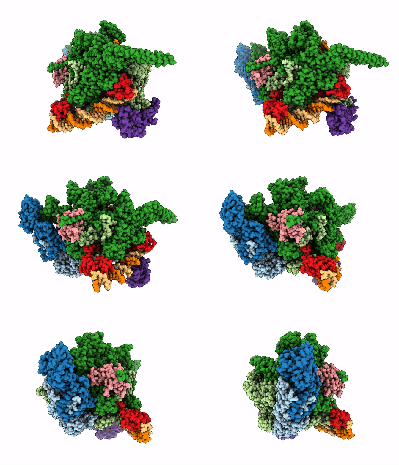
9M9D image
Deposition Date 2025-03-13
Release Date 2026-01-14
Last Version Date 2026-04-15
Entry Detail
PDB ID:
9M9D
Title:
Structural Basis of Pausing During Transcription Initiation in Mycobacterium tuberculosis
Biological Source:
Source Organism(s):
Expression System(s):
Method Details:
Experimental Method:
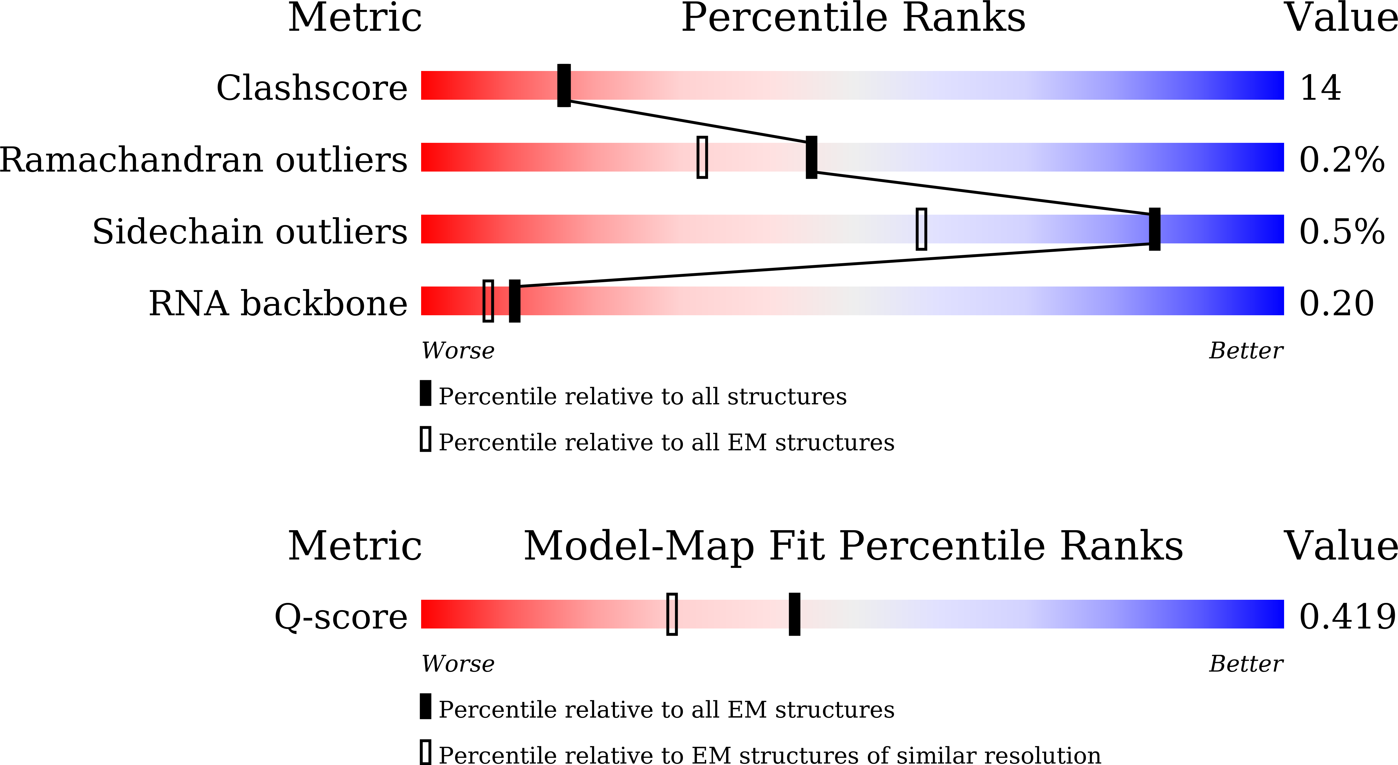
Resolution:
3.29 Å
Aggregation State:
PARTICLE
Reconstruction Method:
SINGLE PARTICLE
Macromolecular Entities
Structures with similar UniProt ID
Protein Blast
Polymer Type:polypeptide(L)
Molecule:DNA-directed RNA polymerase s
Gene (Uniprot):rpoA
Chain IDs:A, B
Chain Length:347
Number of Molecules:2
Biological Source:Mycobacterium tuberculosis H37Rv
Structures with similar UniProt ID
Protein Blast
Polymer Type:polypeptide(L)
Molecule:DNA-directed RNA polymerase s
Gene (Uniprot):rpoB
Chain IDs:C
Chain Length:1178
Number of Molecules:1
Biological Source:Mycobacterium tuberculosis H37Rv
Structures with similar UniProt ID
Protein Blast
Polymer Type:polypeptide(L)
Molecule:DNA-directed RNA polymerase s
Gene (Uniprot):rpoC
Chain IDs:D
Chain Length:1326
Number of Molecules:1
Biological Source:Mycobacterium tuberculosis H37Rv
Structures with similar UniProt ID
Protein Blast
Polymer Type:polypeptide(L)
Molecule:DNA-directed RNA polymerase s
Gene (Uniprot):rpoZ
Chain IDs:E
Chain Length:110
Number of Molecules:1
Biological Source:Mycobacterium tuberculosis H37Rv
Structures with similar UniProt ID
Protein Blast
Polymer Type:polypeptide(L)
Molecule:ECF RNA polymerase sigma fact
Gene (Uniprot):sigE
Chain IDs:F
Chain Length:257
Number of Molecules:1
Biological Source:Mycobacterium tuberculosis H37Rv
Polymer Type:polydeoxyribonucleotide
Molecule:DNA (48-MER)
Chain IDs:H (auth: G)
Chain Length:48
Number of Molecules:1
Biological Source:Mycobacterium tuberculosis H37Rv
Polymer Type:polydeoxyribonucleotide
Molecule:DNA (48-MER)
Chain IDs:I (auth: H)
Chain Length:48
Number of Molecules:1
Biological Source:Mycobacterium tuberculosis H37Rv
Polymer Type:polyribonucleotide
Molecule:RNA (5'-R(*CP*CP*CP*UP*CP*GP*
Chain IDs:J (auth: I)
Chain Length:7
Number of Molecules:1
Biological Source:Mycobacterium tuberculosis H37Rv
Structures with similar UniProt ID
Protein Blast
Polymer Type:polypeptide(L)
Molecule:RNA polymerase-binding transc
Gene (Uniprot):carD
Chain IDs:G (auth: M)
Chain Length:169
Number of Molecules:1
Biological Source:Mycobacterium tuberculosis H37Rv
Primary Citation
Structural basis of pausing during transcription initiation in mycobacterium tuberculosis.
Nat Commun 17 ? ? (2026)
PMID: 41617697 DOI: 10.1038/s41467-026-69104-w

Abstact

In bacteria, RNA polymerase (RNAP) often pauses during the early stages of transcription initiation. The structural basis for these transient pauses remains unclear. Here, we present cryo-electron microscopy (cryo-EM) structures of the paused initiation complex (PIC) and initiation complex (IC) of Mycobacterium tuberculosis (Mtb), which include the RNAP core enzyme, the ECF sigma factor sigma(E), transcription factor CarD, promoter DNA, and nascent RNA. Our structures with pre-melted scaffolds reveal an intermediate at the 6-7 nt stage compatible with a paused-like intermediate, associated with steric hindrance between the emerging RNA and the sigma3.2 region. This clash triggers a swivel of the RNAP structural module and scrunching of the transcription bubble. We also observe positional rearrangement of the sigma4 domain, suggesting a poised pre-escape state. In addition, complementary reconstructions with fully matched DNA scaffolds (N-IC and N-PIC) support the physiological relevance of the captured intermediates. Together, our results support the existence of a mechanistic checkpoint during transcription initiation and suggest an RNA-induced model how RNAP conformational dynamics regulate early transcription.

Legend

Protein

Chemical

Disease

Primary Citation of related structures
Feedback Form
Name
Email
Institute
Feedback