

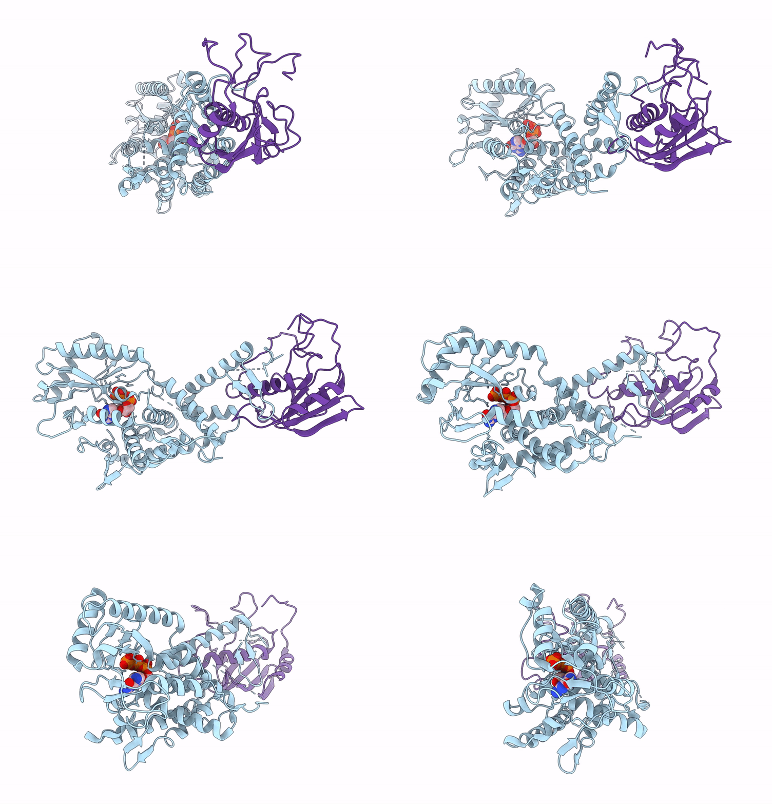
9KKB image
Deposition Date 2024-11-13
Release Date 2025-11-19
Last Version Date 2026-03-18
Entry Detail
PDB ID:
9KKB
Title:
Cryo-EM Structure of CdnG-E2 complex with GTP from Serratia marcescens
Biological Source:
Source Organism(s):
Expression System(s):
Method Details:
Experimental Method:
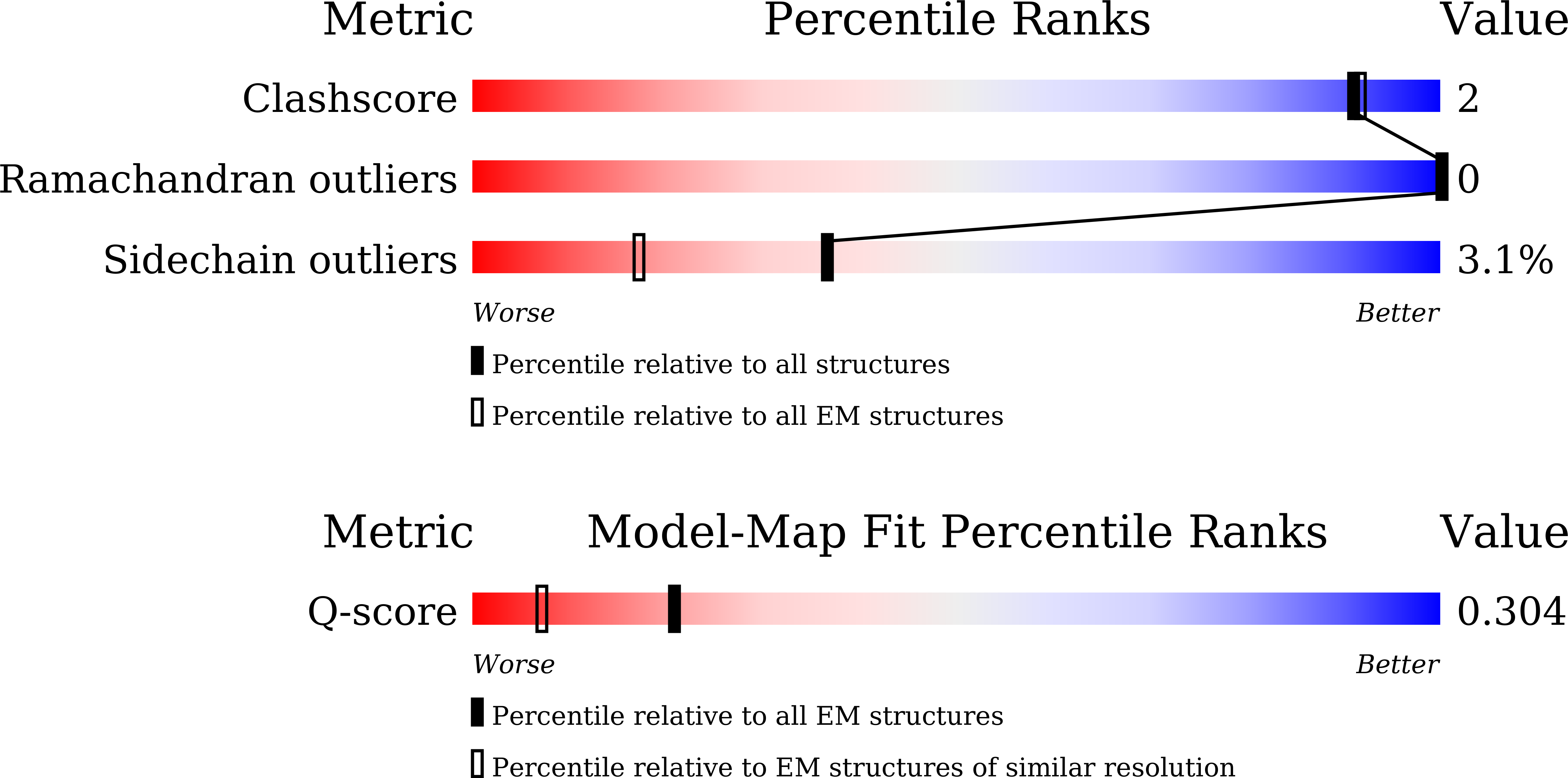
Resolution:
3.36 Å
Aggregation State:
PARTICLE
Reconstruction Method:
SINGLE PARTICLE
Macromolecular Entities
Protein Blast
Polymer Type:polypeptide(L)
Molecule:CdnG
Chain IDs:A
Chain Length:407
Number of Molecules:1
Biological Source:Serratia marcescens
Protein Blast
Polymer Type:polypeptide(L)
Molecule:E2
Chain IDs:B
Chain Length:162
Number of Molecules:1
Biological Source:Serratia marcescens
Ligand Molecules
Primary Citation
Structures and mechanism of E2-CBASS anti-phage system.
Mlife 5 99 107 (2026)
PMID: 41767953 DOI: 10.1002/mlf2.70052

Abstact

Bacteria deploy diverse innate immune systems to combat bacteriophage infections. The cyclic-oligonucleotide-based anti-phage signaling system (CBASS) is a type of innate prokaryotic immune system. CBASS synthesizes cyclic-oligonucleotide through cGAS/DncV-like nucleotidyltransferases (CD-NTases) to activate downstream effectors, which kill bacteriophage-infected bacteria, thereby stopping phage spread. One major class of CBASS contains a homolog of eukaryotic ubiquitin-conjugating enzymes, either as an E1-E2 fusion or a single E2 enzyme. Both enzymes function by regulating CD-NTase activity. Currently, many structures of CD-NTases have been reported, but there are only a few reports of structures where CD-NTases form complexes with the associated E2. In this study, we analyzed the length and classification of the CD-NTase in two types of type II CBASS-E1E2/JAB-CBASS and E2-CBASS. We found that the CD-NTase in E2-CBASS is longer and predominantly belongs to clade G. We also present the structure of the SmCdnG-SmE2 complex with the bound GTP substrate, which indicates the conservation of the donor binding pattern. Interestingly, we discovered that SmCdnG contains a conserved C-terminal alpha-helix and beta-sheet structure, which is uniquely involved in forming a complex with SmE2. We also found that the structure of the E2 protein in the E2-CBASS system is highly conserved. Altogether, we provide mechanistic insights into the E2-CBASS system.

Legend

Protein

Chemical

Disease

Primary Citation of related structures
Feedback Form
Name
Email
Institute
Feedback