

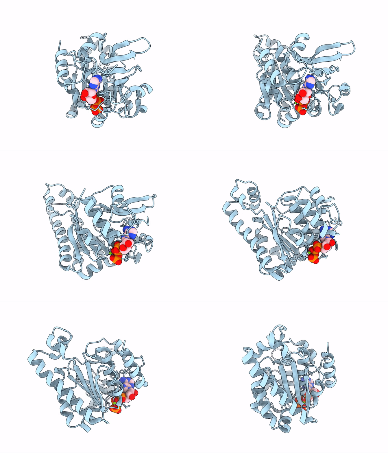
9JV8 image
Deposition Date 2024-10-08
Release Date 2025-10-08
Last Version Date 2026-04-22
Entry Detail
PDB ID:
9JV8
Title:
Crystal structure of M. tuberculosis EccCb1-D2 domain
Biological Source:
Source Organism(s):
Expression System(s):
Method Details:
Experimental Method:
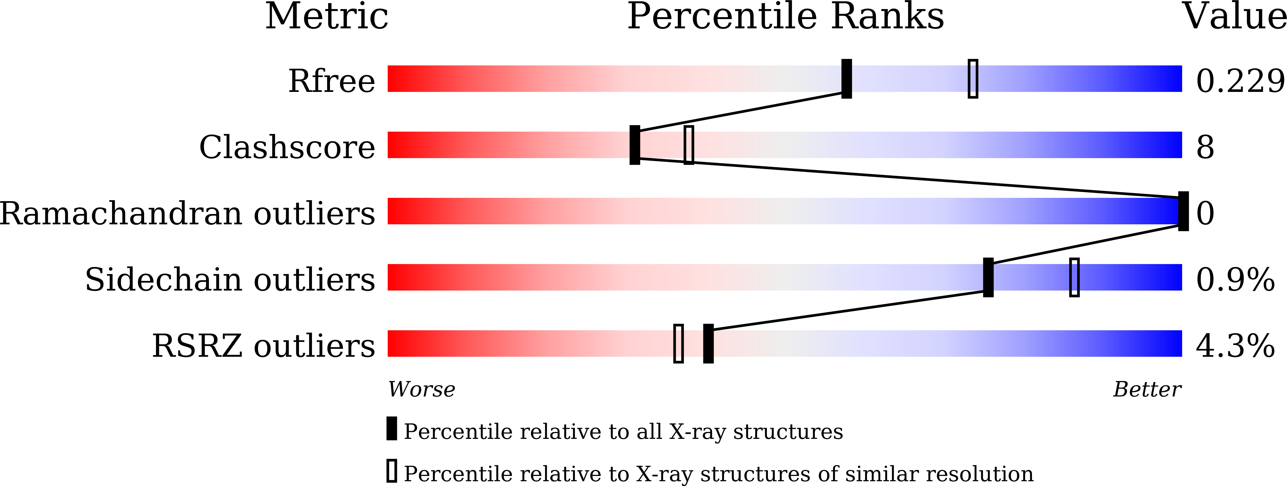
Resolution:
2.20 Å
R-Value Free:
0.22
R-Value Work:
0.17
R-Value Observed:
0.18
Space Group:
P 21 21 21
Macromolecular Entities
Structures with similar UniProt ID
Protein Blast
Polymer Type:polypeptide(L)
Molecule:ESX-1 secretion system protei
Gene (Uniprot):eccCb1
Chain IDs:A
Chain Length:312
Number of Molecules:1
Biological Source:Mycobacterium tuberculosis H37Rv
Primary Citation
Structural analysis of M. tuberculosis EccC1 and its complex with EsxAB virulence factor using X-ray crystallography, molecular docking, and dynamics simulation techniques.
Int. J. Biol. Macromol. 319 145279 145279 (2025)
PMID: 40541887 DOI: 10.1016/j.ijbiomac.2025.145279

Abstact

M. tuberculosis ESX-1 system secretes virulence factors into host macrophages during infection, however, the mechanism of secretion is currently unknown. Here, we have determined the crystal structure of MtbEccCb1-D2 protein (Leu34-Ser313 residues, Mw approximately 31.4 kDa) in complex with ATPgammaS and Mg(2+), which adopts a classical Ftsk/SpoEIII type fold. The EccCb1-D2 showed two melting temperatures, Tm1 at 37.64 +/- 0.08 degrees C and Tm2 at 65.85 +/- 0.12 degrees C, during the unfolding pathway. Modeled ∆EccC1 and ∆EccC1 + EsxAB hexamers showed a channel ( approximately 34 A) involved in EsxAB ( approximately 29 A) translocation toward the inner membrane. At the entrance gate of the channel, the LxxxMxF motif of the EsxB export arm binds to the substrate binding pocket of the EccCb1-D3 protein. Inside the channel, the PL-1 and PL-2 pore loops, close to the alpha7-helix and the loop between beta8-beta9 strands in EccCa1-D1, EccCb1-D2, and EccCb1-D3 may be involved in EsxAB factor translocation. Stability, fluctuation, and compactness parameters in 100 ns dynamics simulation analysis showed the highest flexibility in DeltaEccCa1, ∆EccC1, and ∆EccC1 + EsxAB hexamers and stability in DeltaEccCb1 hexamer. Our EccCb1-D2 structure and dynamics simulation analysis on four modeled systems have revealed the mechanism involved in EsxAB translocation, a key target for the development of antivirulence inhibitors against M. tuberculosis.

Legend

Protein

Chemical

Disease

Primary Citation of related structures
Feedback Form
Name
Email
Institute
Feedback