Deposition Date
2024-10-04
Release Date
2025-10-22
Last Version Date
2026-05-06
Entry Detail
PDB ID:
9JTD
Keywords:
Title:
Crystal structure of PCoV-GD receptor binding domain complexed with fox ACE2
Biological Source:
Source Organism(s):
Vulpes vulpes (Taxon ID: 9627)
Pangolin coronavirus (Taxon ID: 2708335)
Pangolin coronavirus (Taxon ID: 2708335)
Expression System(s):
Method Details:
Experimental Method:
Resolution:
3.59 Å
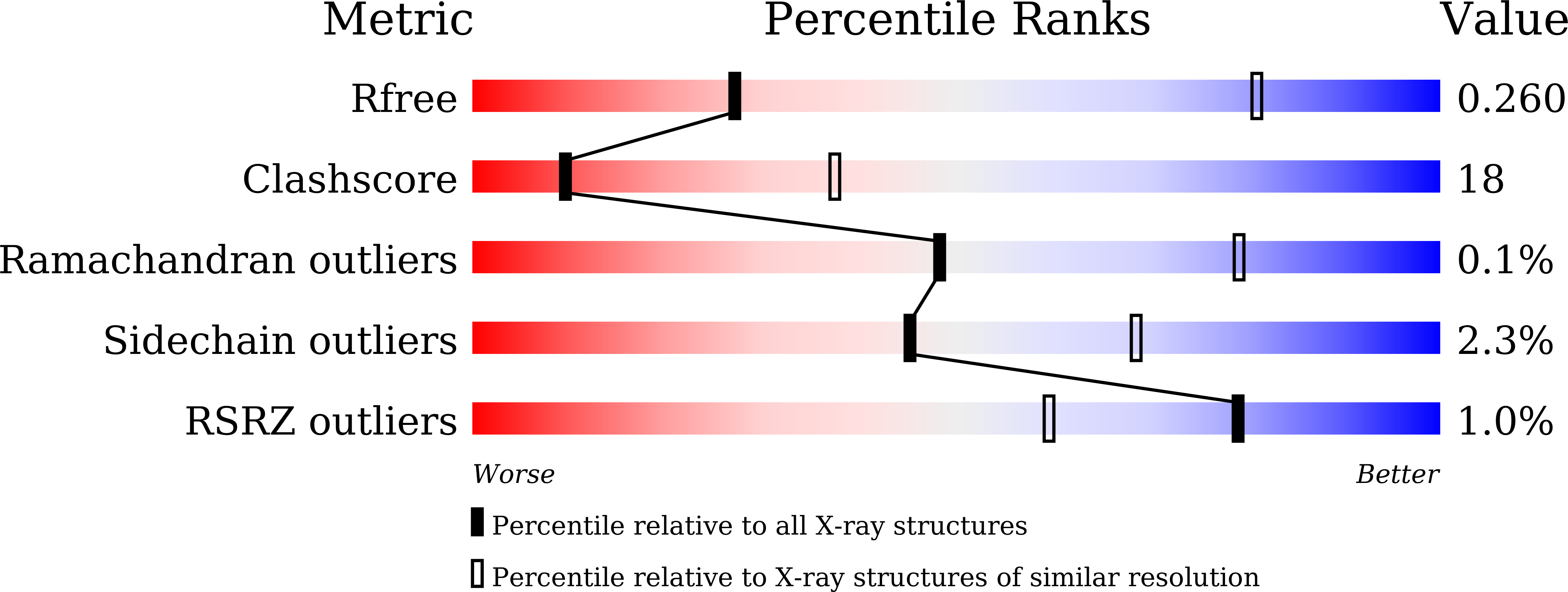
R-Value Free:
0.26
R-Value Work:
0.22
R-Value Observed:
0.23
Space Group:
I 41 2 2