Deposition Date
2024-09-17
Release Date
2026-04-22
Last Version Date
2026-04-22
Entry Detail
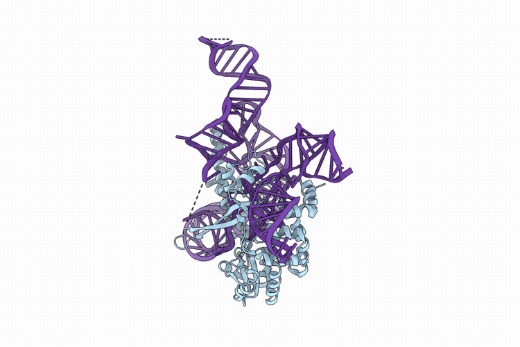
Biological Source:
Source Organism(s):
Klebsiella pneumoniae (Taxon ID: 573)
Expression System(s):
Method Details:
Experimental Method:
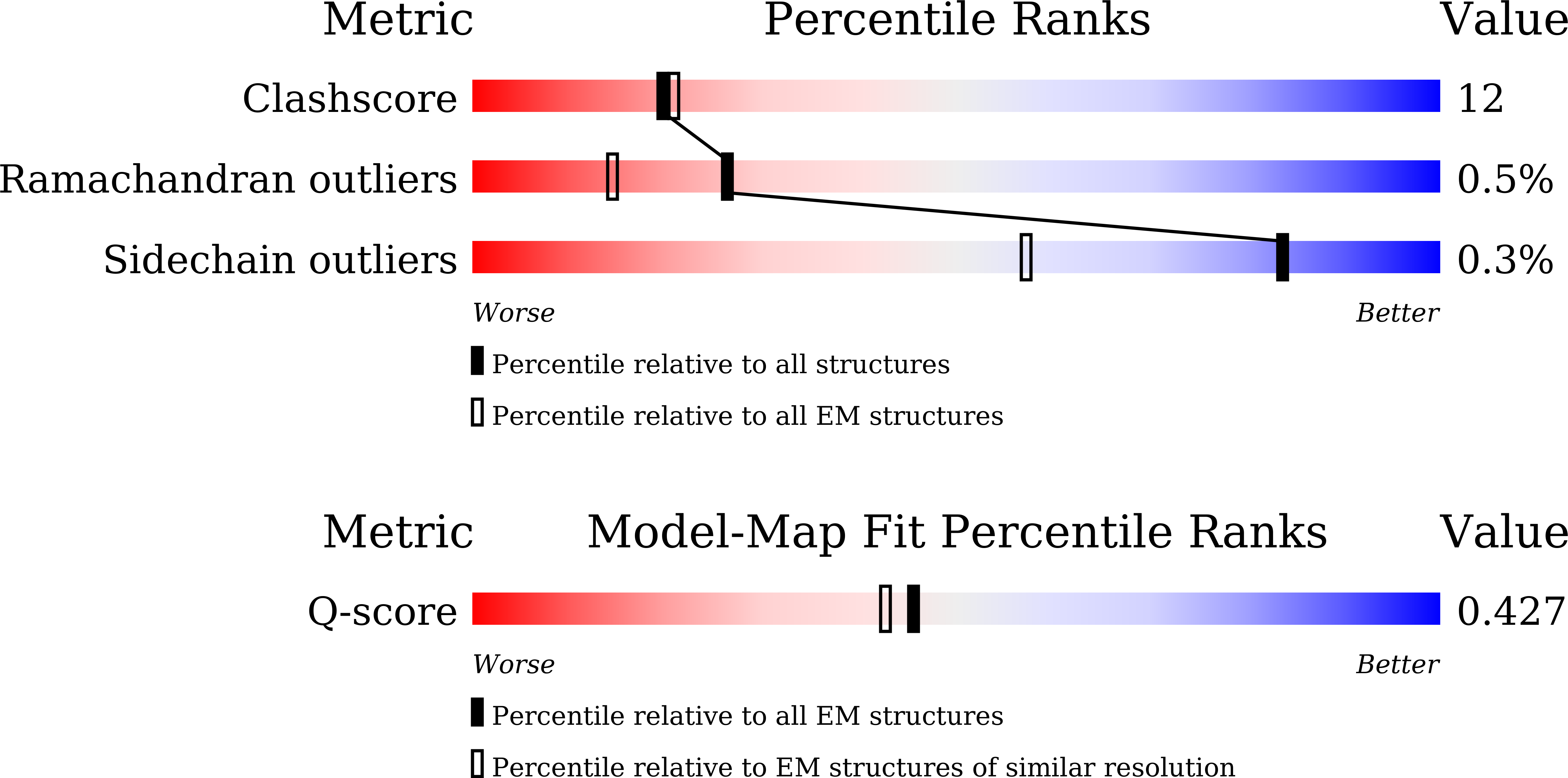
Resolution:
3.49 Å
Aggregation State:
PARTICLE
Reconstruction Method:
SINGLE PARTICLE