9JHQ image
Deposition Date 2024-09-10
Release Date 2025-09-10
Last Version Date 2026-03-04
Entry Detail
PDB ID:
9JHQ
Keywords:
Title:
Crystal structure of GodF, a post-translational modification enzyme involved in the biosynthesis of goadsporin
Biological Source:
Source Organism(s):
Expression System(s):
Method Details:
Experimental Method:
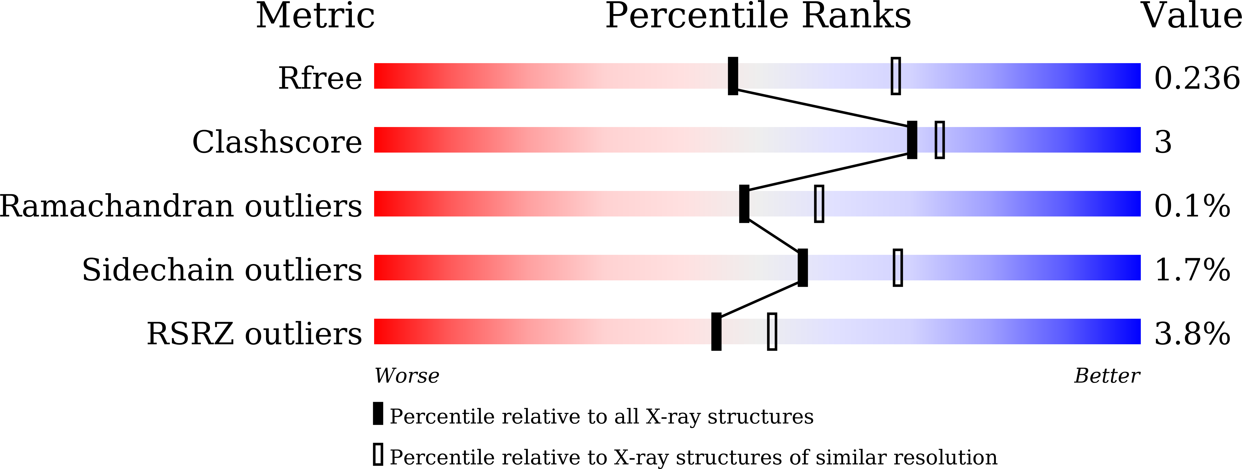
Resolution:
2.34 Å
R-Value Free:
0.23
R-Value Work:
0.18
R-Value Observed:
0.18
Space Group:
C 1 2 1
Macromolecular Entities
Structures with similar UniProt ID
Protein Blast
Polymer Type:polypeptide(L)
Molecule:Goadsporin biosynthetic prote
Gene (Uniprot):godF
Chain IDs:A
Chain Length:867
Number of Molecules:1
Biological Source:Streptomyces sp. TP-A0584
Primary Citation
Structural Analysis of GodF, an O-glutamylation Enzyme Involved in Goadsporin Biosynthesis.
J. Mol. Biol. 438 169619 169619 (2026)
PMID: 41478604 DOI: 10.1016/j.jmb.2025.169619

Abstact

Goadsporin is one of linear azole-containing peptides (LAPs) that form a subgroup within ribosomally synthesized and post-translationally modified peptides (RiPPs). It contains two dehydroalanine residues formed through the action of two enzymes, GodF and GodG, in a two-step process involving serine O-glutamylation followed by elimination. Here, we report the X-ray crystal structure of GodF, which catalyzes the tRNA-dependent glutamylation of target serine residues, resolved at a 2.34-A resolution. Although GodF exhibits low homology at the primary sequence level, its overall structure closely resembles that of TbtB, a tRNA(Glu)-dependent enzyme involved in thiopeptide biosynthesis, as well as the O-glutamylation domains of NisB and MibB, which serve as dehydroalanine synthases in lanthipeptide biosynthesis. The residues and structural elements forming the active site are well-aligned among these enzymes, while regions outside the active site are poorly conserved. Like TbtB, GodF features a coiled-coil subdomain at its N-terminus, and AlphaFold3 predicts this region plays a key role in recognizing the substrate tRNA(Glu). GodF also contains a typical RiPP recognition element (RRE) motif; however, the spatial arrangement of the secondary structural elements comprising this motif differs notably from those in other O-glutamylating enzymes. These structural characteristics of GodF highlight the diversity of substrate-binding pockets among RiPP-modifying enzymes, reflecting the variability in their substrate peptides and the necessity to accommodate distinct conformational and physicochemical properties.

Legend

Protein

Chemical

Disease

Primary Citation of related structures
Feedback Form
Name
Email
Institute
Feedback