

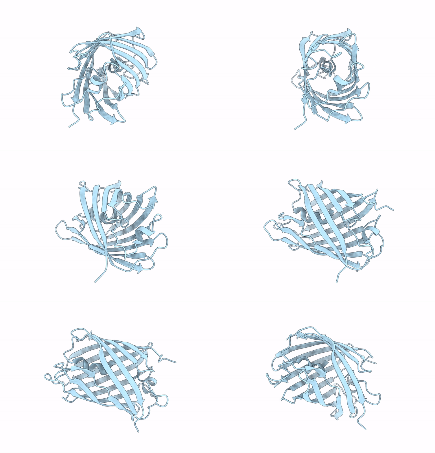
9IRJ image
Deposition Date 2024-07-16
Release Date 2025-07-09
Last Version Date 2026-09-23
Entry Detail
PDB ID:
9IRJ
Title:
X-ray structure of PSmOrange3 (trigonal form)
Biological Source:
Source Organism(s):
Discosoma sp. (Taxon ID: 86600)
Expression System(s):
Method Details:
Experimental Method:
Resolution:
2.80 Å
R-Value Free:
0.20
R-Value Work:
0.17
Space Group:
P 32 2 1
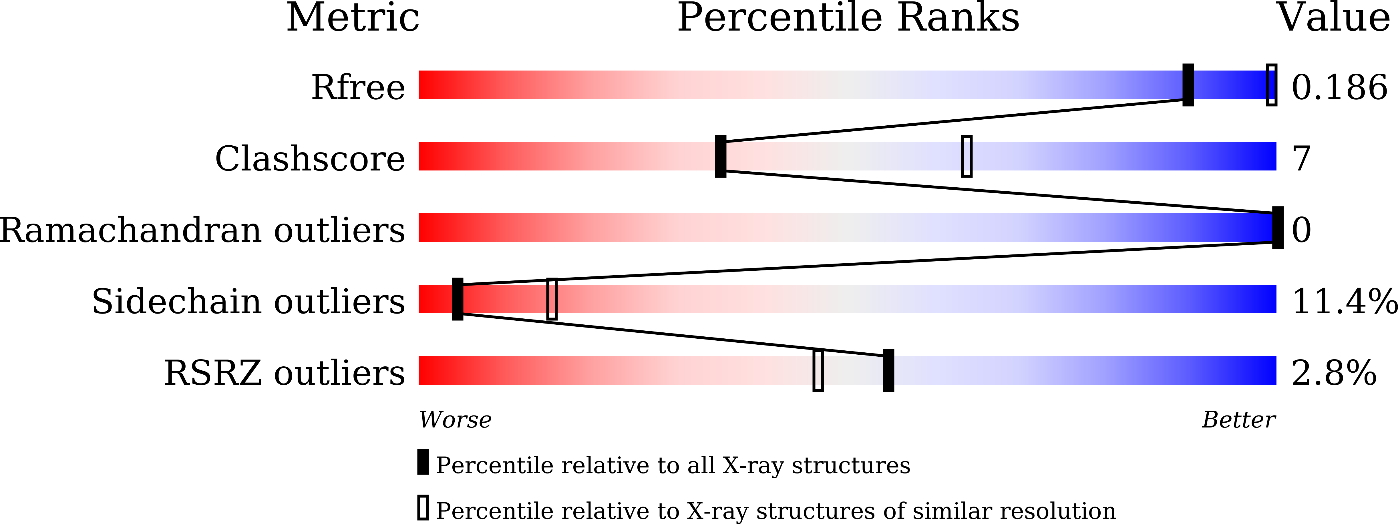
Macromolecular Entities
Polymer Type:polypeptide(L)
Molecule:mOrange
Chain IDs:A
Chain Length:235
Number of Molecules:1
Biological Source:Discosoma sp.
Modified Residue
Compound ID Chain ID Parent Comp ID Details 2D Image
CRO A GLY chromophore
Primary Citation
Next-generation orange-to-far-red photoconvertible fluorescent protein for single-molecule microscopy and protein dynamic tracking.
Sci Rep 15 40166 40166 (2025)
PMID: 41249261 DOI: 10.1038/s41598-025-23873-4

Abstact

The PSmOrange and PSmOrange2 fluorescent proteins undergo irreversible photoconversion from the orange to far-red form under blue light, which makes them probes of choice for protein tracking and single-molecule super-resolution imaging. However, both proteins exhibit noticeable photoconversion under 550-570 nm light used for excitation of their orange form, which complicates applications of these photoconvertible FPs in cell imaging experiments. Here, we report the next-generation PSmOrange variant, called PSmOrange3, which is characterized by minimal photoconversion under 550-570 nm light and high photoconversion contrast. PSmOrange3 undergoes efficient photoconversion from the orange (Ex/Em at 550 nm/564 nm) to far-red form (Ex/Em at 614 nm/655 nm) with 430-470 nm violet-blue light of moderate power density (3-180 mW/mm(2)) in a native cellular environment. The molecular brightness of orange and far-red forms of PSmOrange3 was 1.2- and 1.4-fold brighter than that of PSmOrange2. In addition, PSmOrange3 had a substantially higher photostability of the orange form but a little less photostability of the far-red form. We solved the crystal structure of PSmOrange3 at a 2.8 A resolution, which confirmed its monomeric state and revealed the role of the introduced mutations in the properties of PSmOrange3. Using mass spectrometry we revealed the chemical structure of the PSmOrange3 chromophore before and after photoconversion. PSmOrange3 was properly localized with different protein fusions and photoconverted from the orange to far-red state inside live and fixed mammalian cells without exogenously supplied oxidants. Among all proteins of the PSmOrange series, both forms of PSmOrange3 were the brightest in the reducing environment of the mitochondrial lumen. PSmOrange3 photoconverted efficiently with blue light and almost did not photoconvert with green light, which allows investigators to excite its orange form and photoconvert it to the far-red form with different light. We demonstrated the applicability of PSmOrange3 for photoactivated localization microscopy (PALM) of tubulin microtubules using 488-nm photoconversion, achieving mean localization precision per single-molecule event of 24.6 and 23.3 nm in fixed and live mammalian cells, respectively. We believe that PSmOrange3 can represent a suitable alternative to the PSmOrange and PSmOrange2 proteins and will be a valuable addition to the repertoire of available photoconvertible fluorescent proteins.

Legend

Protein

Chemical

Disease

Primary Citation of related structures
Feedback Form
Name
Email
Institute
Feedback