

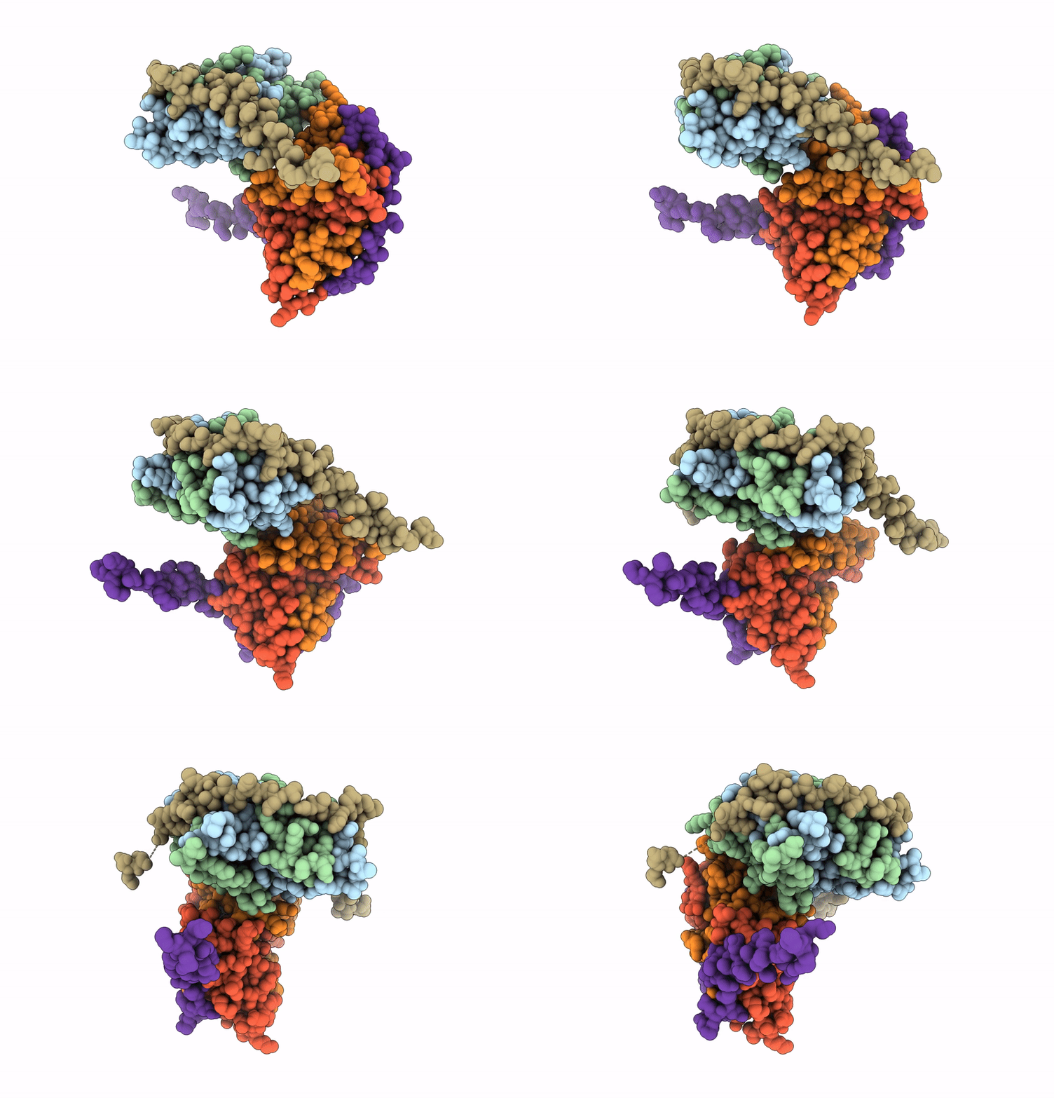
9INC image
Deposition Date 2024-07-06
Release Date 2025-07-09
Last Version Date 2026-07-22
Entry Detail
PDB ID:
9INC
Keywords:
Title:
High resolutional Crystal Structure of human H2A.Z-H2B dimer in complex with human YL1-Z domain
Biological Source:
Source Organism(s):
Homo sapiens (Taxon ID: 9606)
Expression System(s):
Method Details:
Experimental Method:
Resolution:
2.01 Å
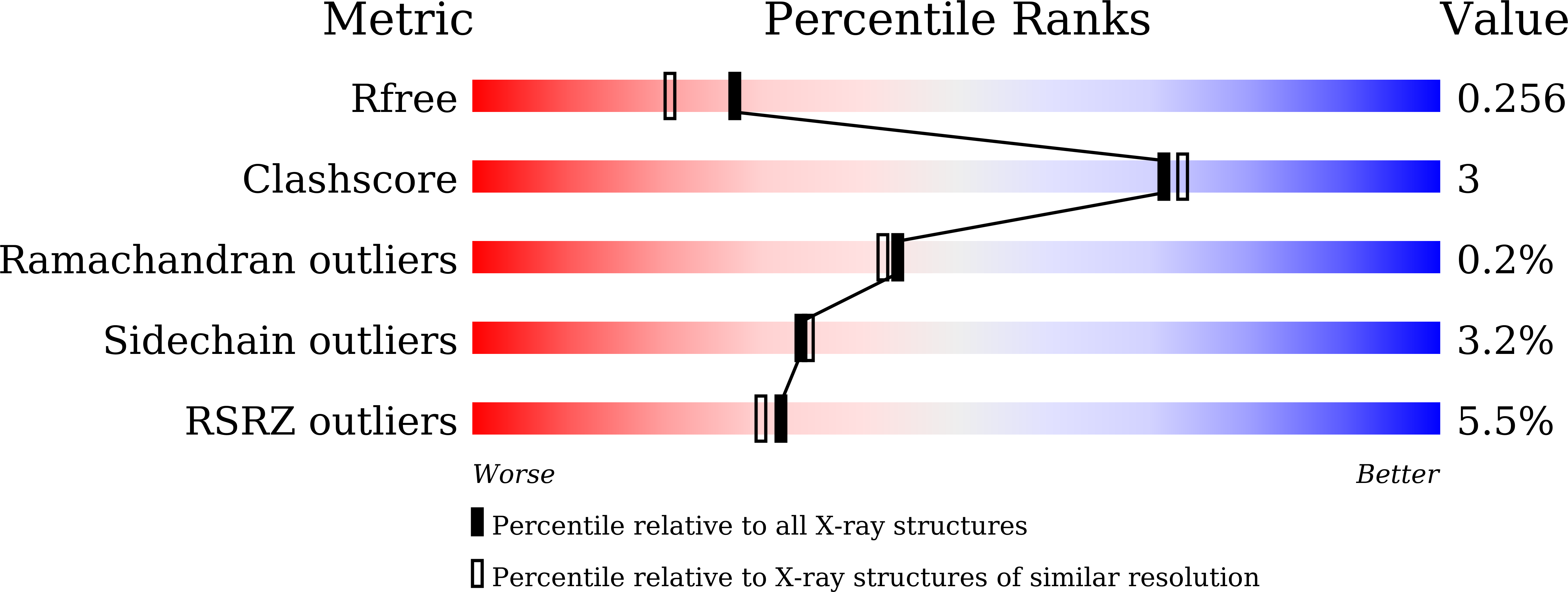
R-Value Free:
0.25
R-Value Work:
0.21
R-Value Observed:
0.22
Space Group:
C 2 2 21
Macromolecular Entities
Polymer Type:polypeptide(L)
Molecule:Histone H2A.Z
Gene (Uniprot):H2AZ1
Chain IDs:A, D
Chain Length:98
Number of Molecules:2
Biological Source:Homo sapiens
Polymer Type:polypeptide(L)
Molecule:Histone H2B type 1-J
Gene (Uniprot):H2BC11
Chain IDs:B, E
Chain Length:94
Number of Molecules:2
Biological Source:Homo sapiens
Polymer Type:polypeptide(L)
Molecule:Vacuolar protein sorting-asso
Gene (Uniprot):VPS72
Chain IDs:C, F
Chain Length:87
Number of Molecules:2
Biological Source:Homo sapiens
Primary Citation
Structural insights into specific recognition of the histone variant H2A.Z by the YL1 subunit of SRCAP chromatin-remodeling complex.
J.Biol.Chem. 302 111294 111294 (2026)
PMID: 41708003 DOI: 10.1016/j.jbc.2026.111294

Abstact

The SNF2-related CREB-binding protein activator protein complex regulates gene transcription through chromatin remodeling, where the subunit YL1 mediates the exchange of H2A-H2B dimers with H2A.Z-H2B in the nucleosome. Although structures of YL1 in complex with H2A.Z-H2B have been previously reported for both Drosophila and Homo sapiens, the complex structures of the two species exhibit significant differences, which have caused some confusion over how YL1 recruits H2A.Z-H2B. We determined the high-resolution (2.01 A) crystal structure of the N-terminal region (8-73) of the human SNF2-related CREB-binding protein activator protein subunit YL1 in complex with H2B-H2A.Z; this domain is termed as YL1-Z. YL1-Z consists of an alpha helix and a longer loop, which bind to H2A.Z-H2B to form a complex. In the structure, residues Phe29, Tyr30, Tyr34, and Phe37 of the YL1-Z domain specifically recognize the hydrophobic residues, Gln87, Ile90, Ile100, and Ile104, of H2A.Z through hydrophobic interactions, forming a hydrophobic core. In addition, residues Asp55, Asp58, and Asp60 of YL1-Z form electrostatic interactions with Arg34 and Lys37 on H2A.Z, stabilizing the complex structure. The particular recognition has been validated in vitro by maltose-binding protein-pulldown and isothermal titration calorimetry experiments. Our data show that YL1 employs a conserved structural mechanism for recognizing the H2A.Z-H2B dimer.

Legend

Protein

Chemical

Disease

Primary Citation of related structures
Feedback Form
Name
Email
Institute
Feedback