

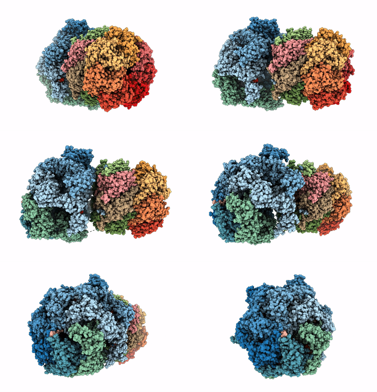
9IF4 image
Deposition Date 2025-02-17
Release Date 2025-08-27
Last Version Date 2026-09-09
Entry Detail
PDB ID:
9IF4
Keywords:
Title:
Structure of the Mycobacterium Tuberculosis ClpC1P1P2 complex bound to the activator Bz-Leu-Leu
Biological Source:
Source Organism(s):
Expression System(s):
Method Details:
Experimental Method:
Resolution:
3.09 Å
Aggregation State:
PARTICLE
Reconstruction Method:
SINGLE PARTICLE

Macromolecular Entities
Polymer Type:polypeptide(L)
Molecule:ATP-dependent Clp protease AT
Gene (Uniprot):clpC1
Chain IDs:A, B, C, D, E, F
Chain Length:658
Number of Molecules:6
Biological Source:Mycobacterium tuberculosis
Polymer Type:polypeptide(L)
Molecule:ATP-dependent Clp protease pr
Gene (Uniprot):clpP2
Chain IDs:G, H, I, J, K (auth: L), L (auth: M), T (auth: K)
Chain Length:200
Number of Molecules:7
Biological Source:Mycobacterium tuberculosis
Polymer Type:polypeptide(L)
Molecule:ATP-dependent Clp protease pr
Gene (Uniprot):clpP1
Chain IDs:M (auth: N), N (auth: O), O (auth: P), P (auth: Q), Q (auth: R), R (auth: S), S (auth: T)
Chain Length:179
Number of Molecules:7
Biological Source:Mycobacterium tuberculosis
Polymer Type:polypeptide(L)
Molecule:activator Bz-Leu-Leu
Chain IDs:U, V, W, X (auth: Y), Y (auth: Z), Z (auth: a), AA (auth: b)
Chain Length:2
Number of Molecules:7
Biological Source:synthetic construct
Polymer Type:polypeptide(L)
Molecule:Unknown peptide
Chain IDs:BA (auth: X)
Chain Length:26
Number of Molecules:1
Biological Source:Mycobacterium tuberculosis
Primary Citation
Activation mechanism and structural assembly of the Mycobacterium tuberculosis ClpP1P2 protease and its associated ATPases.
Cell Rep 45 117400 117400 (2026)
PMID: 42213777 DOI: 10.1016/j.celrep.2026.117400

Abstact

Supramolecular assemblies are fundamental to cellular biochemical processes, relying on their dynamic nature to perform essential functions. The protease ClpP1P2, in association with ATPase partners ClpC1 or ClpX, is critical for the survival of Mycobacterium tuberculosis (Mtb). While the ClpP1P2 complex requires activation by specific N-blocked dipeptides to exhibit proteolytic activity in vitro, the mechanism of in vivo activation remains unclear. In this study, we use cryo-electron microscopy (cryo-EM) to determine the structure of the ClpC1P1P2 complex, revealing a highly asymmetric architecture with ClpC1 bound to the ClpP1P2 protease barrel. The activator dipeptide is observed only in the ClpP2 active site, while the ClpP1 entry pore remains closed. Molecular crowding agents promote the formation of larger ClpXP1P2 and ClpC1P1P2 complexes, enhancing structural stability and enzymatic activity. These findings suggest that molecular crowding stabilizes these complexes and promotes their activation, providing new insights into ClpC1P1P2 structural dynamics and function.

Legend

Protein

Chemical

Disease

Primary Citation of related structures
Feedback Form
Name
Email
Institute
Feedback