9IBK image
Deposition Date 2025-02-12
Release Date 2025-11-19
Last Version Date 2026-06-10
Entry Detail
PDB ID:
9IBK
Title:
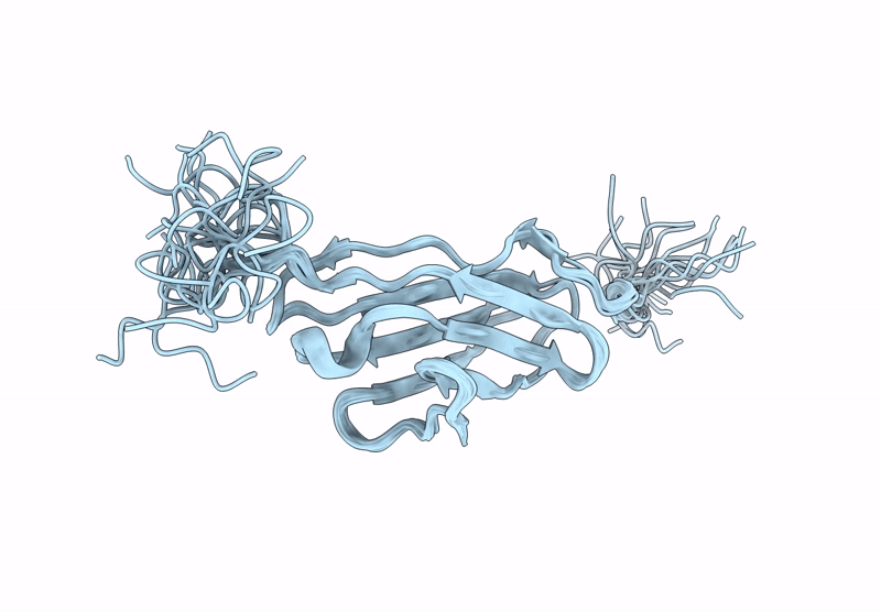
Solution NMR study of the titin I-band IgI domain I82 reveals conformational dynamics
Biological Source:
Source Organism(s):
Mus musculus (Taxon ID: 10090)
Expression System(s):
Method Details:
Experimental Method:
Conformers Calculated:
50
Conformers Submitted:
20
Selection Criteria:
structures with the lowest energy
Macromolecular Entities
Structures with similar UniProt ID
Protein Blast
Polymer Type:polypeptide(L)
Molecule:Titin
Gene (Uniprot):Ttn
Mutagens:n.a.
Chain IDs:A
Chain Length:102
Number of Molecules:1
Biological Source:Mus musculus
Ligand Molecules
Primary Citation
Solution NMR study of the titin I-band IgI domain I82 shows unusual conformational dynamics.
J.Biomol.Nmr 80 ? ? (2026)
PMID: 42223715 DOI: 10.1007/s10858-026-00493-2

Abstact

beta-sandwich domains of the intermediate immunoglobulin fold (IgI) are key building blocks of many large, multidomain proteins - such as titin. The uniformity of the basic fold of these domains does not seem to be an impediment to their adaptation to a wide variety of functions. IgI domains in different regions of titin have to fulfil distinct functions which is seen in subtle differences of sequence, structure and stability. This is particularly true for the N2A region whose IgI domains are subtly distinct from other parts of I-band titin. We have already shown the unusual structure and properties of IgI domain I83 which is able to bind to calcium and is part of a binding site for F-actin and p94. To continue our exploration of the titin IgI domains of the N2A region we describe here the solution NMR structure of domain I82 of murine titin. The structure of the murine domain is virtually identical to the human homologue. However, the NMR investigation reveals the existence of distinct conformers around a highly conserved glycine, part of the tyrosine corner motif at the junction of the EF-loop with the F-strand. (15)N relaxation data show substantial line broadening for residues around this glycine, confirming conformational exchange on the fast-intermediate time scale. The unusual dynamics could for the first time explain the high level of conservation of the tyrosine corner glycine via a function in the folding of the domain.

Legend

Protein

Chemical

Disease

Primary Citation of related structures
Feedback Form
Name
Email
Institute
Feedback