

9IBA image
Deposition Date 2025-02-12
Release Date 2025-12-17
Last Version Date 2026-07-01
Entry Detail
PDB ID:
9IBA
Keywords:
Title:
Crystal structure of Arr-2 in complex with Rifampicin
Biological Source:
Source Organism(s):
Expression System(s):
Method Details:
Experimental Method:
Resolution:
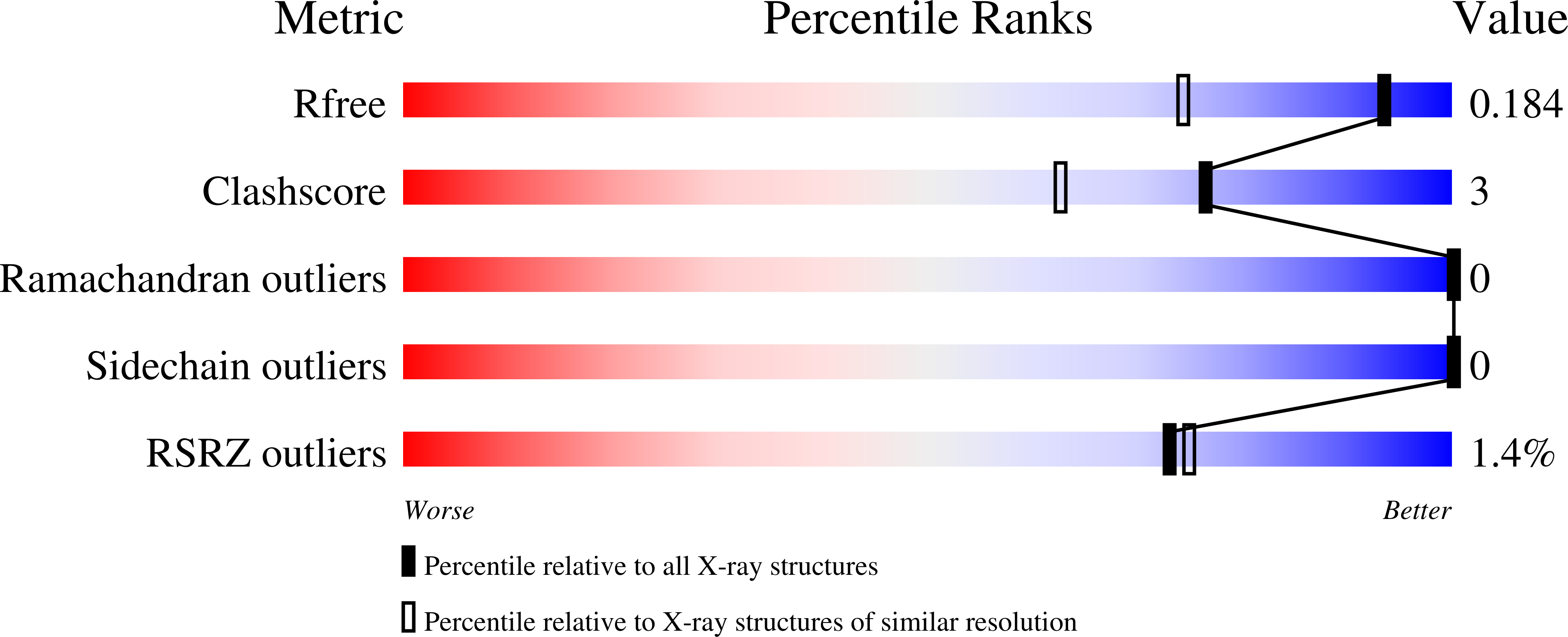
1.40 Å
R-Value Free:
0.18
R-Value Work:
0.14
Space Group:
P 21 21 21
Macromolecular Entities
Polymer Type:polypeptide(L)
Molecule:ARR-2
Gene (Uniprot):arr-2
Chain IDs:A
Chain Length:151
Number of Molecules:1
Biological Source:Pseudomonas aeruginosa
Ligand Molecules
Primary Citation
Discovery of Inhibitors for Bacterial Arr Enzymes ADP-Ribosylating and Inactivating Rifamycin Antibiotics.
Acs Chem.Biol. 20 1584 1593 (2025)
PMID: 40509881 DOI: 10.1021/acschembio.5c00164

Abstact

ADP-ribosylation is an enzymatic process where an ADP-ribose moiety is transferred from NAD(+) to an acceptor molecule. While ADP-ribosylation is well-established as a post-translational modification of proteins, rifamycin antibiotics are its only known small-molecule targets. ADP-ribosylation of rifampicin was first identified in Mycolicibacterium smegmatis, whose Arr enzyme transfers the ADP-ribose moiety to the 23-hydroxy group of rifampicin preventing its interaction with the bacterial RNA polymerase thereby inactivating the antibiotic. Arr homologues are widely spread among bacterial species and present in several pathogenic species often associated with mobile genetic elements. Inhibition of Arr enzymes offers a promising strategy to overcome ADP-ribosylation mediated rifamycin resistance. We developed a high-throughput activity assay which was applied to screen an in-house library of human ADP-ribosyltransferase-targeted compounds. We identified 15 inhibitors with IC(50) values below 5 muM against four Arr enzymes from M. smegmatis, Pseudomonas aeruginosa, Stenotrophomonas maltophilia, and Mycobacteroides abscessus. The observed overall selectivity of the hit compounds over the other homologues indicated structural differences between the proteins. We crystallized M. smegmatis and P. aeruginosa Arr enzymes, the former in complex with its most potent hit compound with an IC(50) value of 1.3 muM. We observed structural differences in the NAD(+) binding pockets of the two Arr homologues explaining the selectivity. Although the Arr inhibitors did not sensitize M. smegmatis to rifampicin in a growth inhibition assay, the structural information and the collection of inhibitors provide a foundation for rational modifications and further development of the compounds.

Legend

Protein

Chemical

Disease

Primary Citation of related structures
Feedback Form
Name
Email
Institute
Feedback