

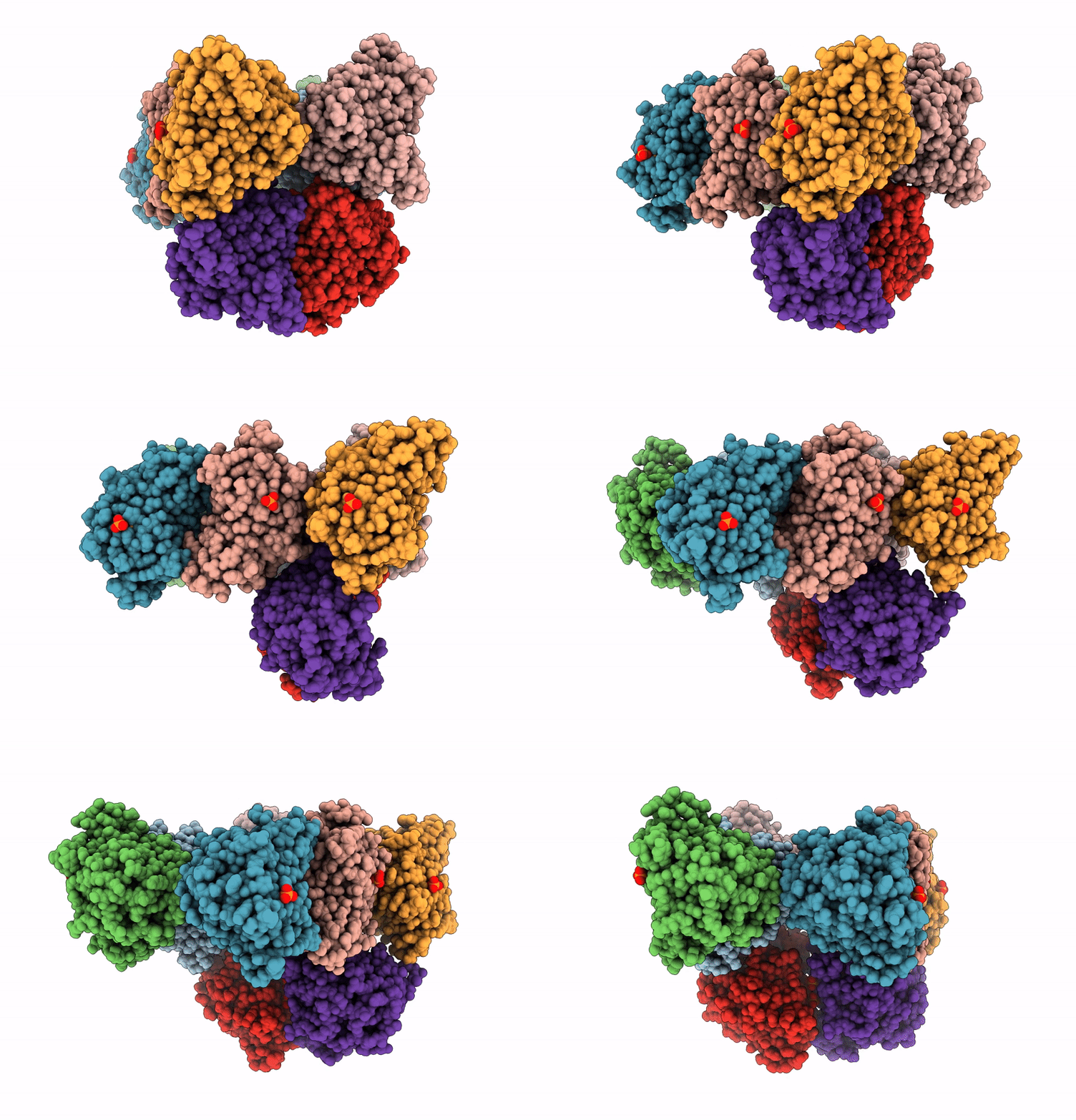
9I1U image
Deposition Date 2025-01-17
Release Date 2025-12-24
Last Version Date 2026-03-04
Entry Detail
PDB ID:
9I1U
Keywords:
Title:
Lytic polysaccharide monooxygenase from Cellvibrio japonicus - CjAA10B
Biological Source:
Source Organism(s):
Expression System(s):
Method Details:
Experimental Method:
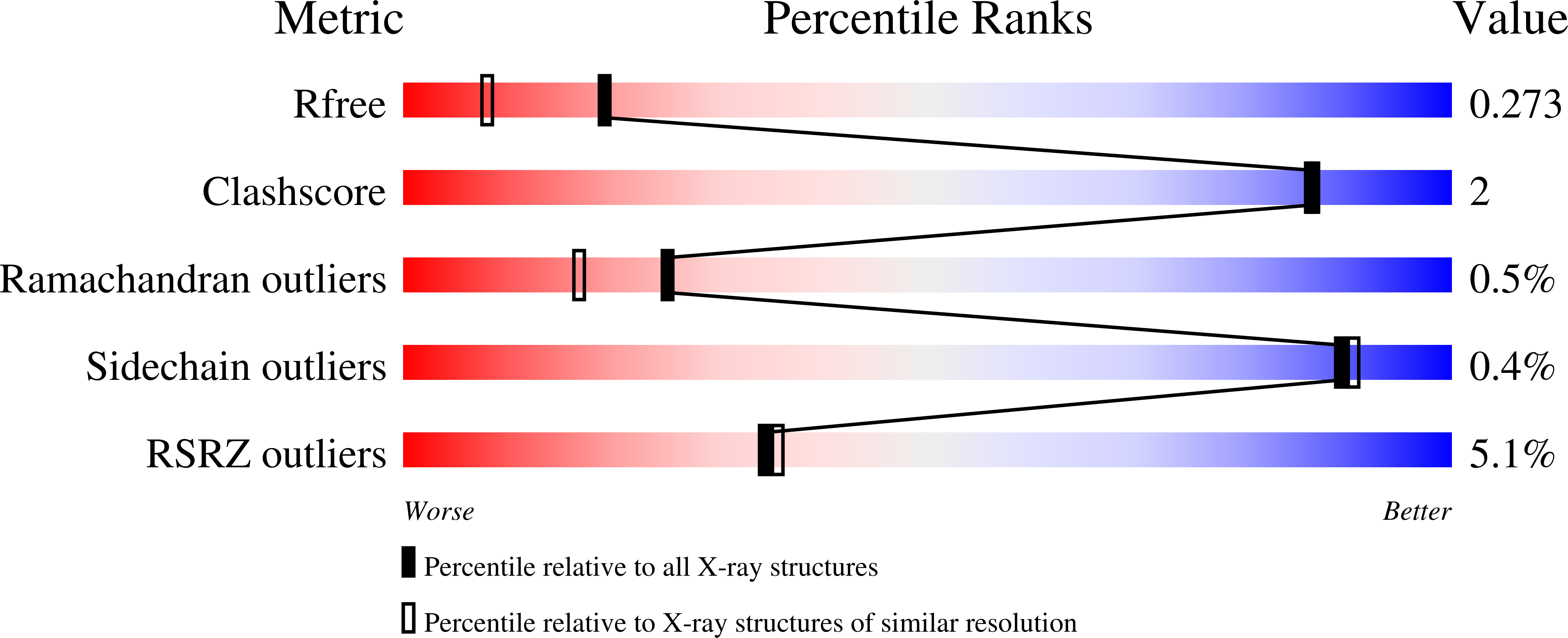
Resolution:
1.90 Å
R-Value Free:
0.27
R-Value Work:
0.23
Space Group:
P 1 21 1
Macromolecular Entities
Structures with similar UniProt ID
Protein Blast
Polymer Type:polypeptide(L)
Molecule:Chitin binding protein, putat
Gene (Uniprot):10B
Chain IDs:A, B, C, D, E, F, G, H
Chain Length:211
Number of Molecules:8
Biological Source:Cellvibrio japonicus
Primary Citation
Electrochemical Insight into the Copper Redox Chemistry and H 2 O 2 and O 2 Reducing Capability of Two AA10 Lytic Polysaccharide Monooxygenases.
Acs Electrochem 2 239 257 (2026)
PMID: 41669087 DOI: 10.1021/acselectrochem.5c00266

Abstact

Lytic polysaccharide monooxygenases ([L]-PMOs) are copper-containing enzymes that catalyse cleavage of the glycosidic bond, a process central to microbial biomass degradation. Here, we describe electrochemical methods used to investigate the Cu(2+/1+) redox chemistry and the polysaccharide-free catalytic activity of two AA10 LPMOs: CjAA10B from Cellvibrio japonicus and CfAA10 from Cellulomonas fimi. Immobilisation of these enzymes on the surface of a graphite electrode allows for direct electrochemical measurements of Cu(2+/1+) redox cycling as well as the ability of both LPMOs to reduce H(2)O(2) vs O(2). These measurements can be advantageous when compared to biological dye assays as they provide direct kinetic measurements and allow for investigation over a wider range of environmental conditions. Values of k (cat) and K (M)- are reported for H(2)O(2) and O(2) reduction by CjAA10B and CfAA10 from pH 5-7, with CfAA10 consistently outperforming CjAA10B. Both enzymes perform faster catalysis with H(2)O(2) but when comparing the affinity-coupled specificity constant (k (cat)/K (M)), the LPMOs perform similarly with both H(2)O(2) and O(2), suggesting both substrates are viable. We also note an increase in redox signals as pH is decreased that correlates with EPR data suggesting a second species is formed <pH 5, postulated to occur due to the protonation of a glutamate residue (pK (a) approximately 4.6). The increase in signal size with decreasing pH that is seen for the non-catalytic Cu(2+/1+) transition is interpreted in light of an increasing proportion of electroactive species at low pH; such a change in activity with pH is notably not observed in the presence of substrate (H(2)O(2) or O(2)). This suggests that substrate binding modulates the active site, disrupting the effect of protonation. These findings establish electrochemistry as a powerful tool for probing LPMO activity.

Legend

Protein

Chemical

Disease

Primary Citation of related structures
Feedback Form
Name
Email
Institute
Feedback