

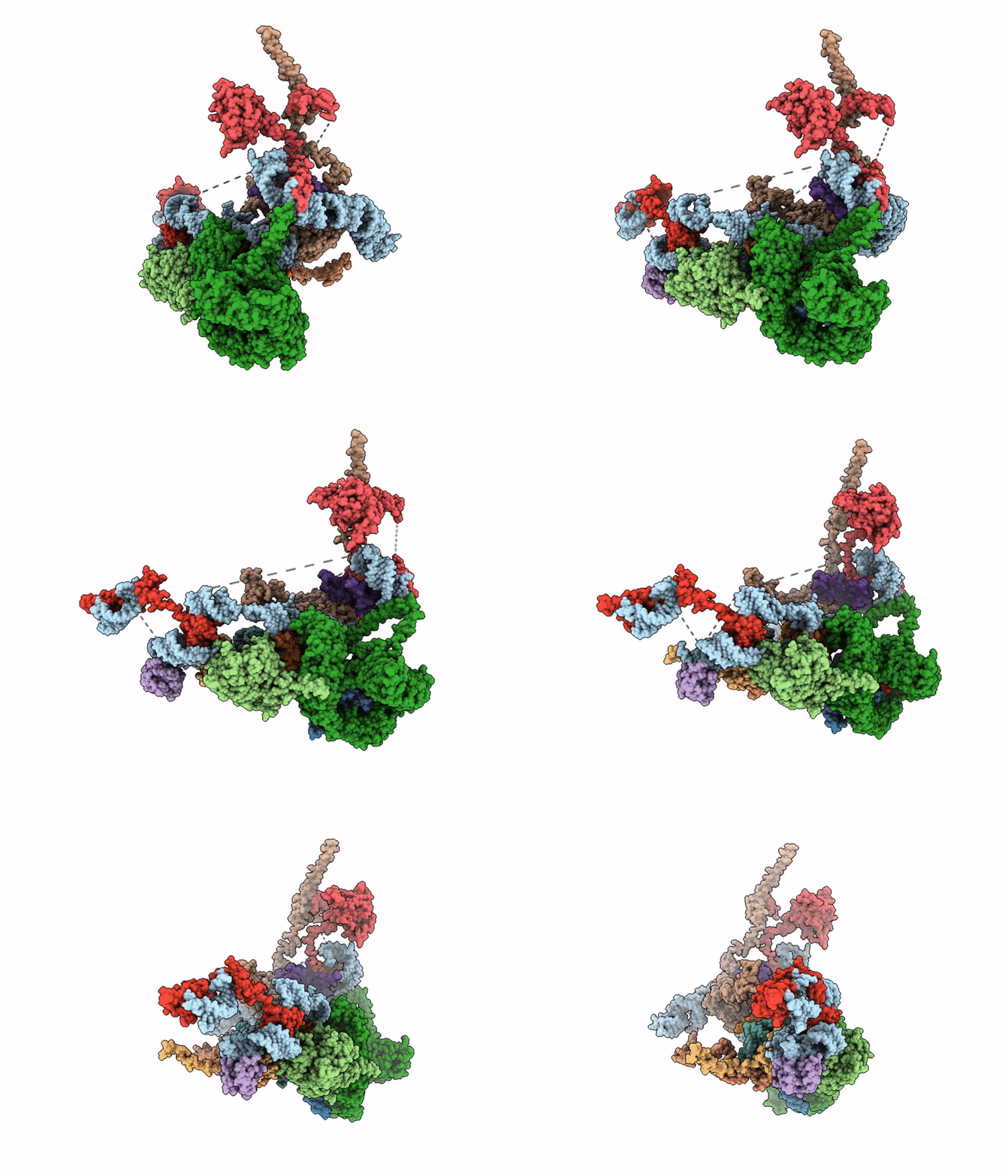
9GJ5 image
Deposition Date 2024-08-21
Release Date 2026-02-18
Last Version Date 2026-06-03
Entry Detail
PDB ID:
9GJ5
Keywords:
Title:
Human 80S ribosome in complex with NatA in distal position and Ebp1
Biological Source:
Source Organism(s):
Homo sapiens (Taxon ID: 9606)
Expression System(s):
Method Details:
Experimental Method:
Resolution:
4.61 Å
Aggregation State:
PARTICLE
Reconstruction Method:
SINGLE PARTICLE
Macromolecular Entities
Polymer Type:polyribonucleotide
Molecule:28S rRNA
Chain IDs:D (auth: 1)
Chain Length:5070
Number of Molecules:1
Biological Source:Homo sapiens
Polymer Type:polypeptide(L)
Molecule:N-alpha-acetyltransferase 10
Gene (Uniprot):NAA10
Chain IDs:A (auth: 2)
Chain Length:171
Number of Molecules:1
Biological Source:Homo sapiens
Polymer Type:polyribonucleotide
Molecule:5.8S rRNA
Chain IDs:B (auth: 8)
Chain Length:157
Number of Molecules:1
Biological Source:Homo sapiens
Polymer Type:polypeptide(L)
Molecule:Proliferation-associated prot
Gene (Uniprot):PA2G4
Chain IDs:M (auth: A)
Chain Length:394
Number of Molecules:1
Biological Source:Homo sapiens
Polymer Type:polypeptide(L)
Molecule:N-alpha-acetyltransferase 15,
Gene (Uniprot):NAA15
Chain IDs:C (auth: B)
Chain Length:840
Number of Molecules:1
Biological Source:Homo sapiens
Polymer Type:polypeptide(L)
Molecule:60S ribosomal protein L4
Gene (Uniprot):RPL4
Chain IDs:E (auth: LC)
Chain Length:427
Number of Molecules:1
Biological Source:Homo sapiens
Polymer Type:polypeptide(L)
Molecule:Large ribosomal subunit prote
Gene (Uniprot):RPL6
Chain IDs:F (auth: LE)
Chain Length:288
Number of Molecules:1
Biological Source:Homo sapiens
Polymer Type:polypeptide(L)
Molecule:60S ribosomal protein L19
Gene (Uniprot):RPL19
Chain IDs:K (auth: LR)
Chain Length:196
Number of Molecules:1
Biological Source:Homo sapiens
Polymer Type:polypeptide(L)
Molecule:60S ribosomal protein L23a
Gene (Uniprot):RPL23A
Chain IDs:J (auth: LX)
Chain Length:156
Number of Molecules:1
Biological Source:Homo sapiens
Polymer Type:polypeptide(L)
Molecule:Large ribosomal subunit prote
Gene (Uniprot):RPL26
Chain IDs:H (auth: LY)
Chain Length:144
Number of Molecules:1
Biological Source:Homo sapiens
Polymer Type:polypeptide(L)
Molecule:60S ribosomal protein L35
Gene (Uniprot):RPL35
Chain IDs:I (auth: Lh)
Chain Length:122
Number of Molecules:1
Biological Source:Homo sapiens
Polymer Type:polypeptide(L)
Molecule:60S ribosomal protein L38
Gene (Uniprot):RPL38
Chain IDs:G (auth: Lk)
Chain Length:70
Number of Molecules:1
Biological Source:Homo sapiens
Polymer Type:polypeptide(L)
Molecule:60S ribosomal protein L28
Gene (Uniprot):RPL28
Chain IDs:L (auth: Lr)
Chain Length:196
Number of Molecules:1
Biological Source:Homo sapiens
Ligand Molecules
Primary Citation
NatA engages in multi-factor complexes at the ribosomal polypeptide tunnel exit.
Nat Commun 17 884 884 (2026)
PMID: 41577663 DOI: 10.1038/s41467-026-68787-5

Abstact

N-terminal acetylation (NTA) is the most common protein modification in eukaryotes, playing a crucial role in proteostasis. Almost 40% of the human proteome is acetylated co-translationally by the NatA complex, which requires prior N-terminal methionine excision (NME). Recently, NatA was shown to form multi-enzyme complexes with MAP1/NAC or MAP2, combining the capabilities of NME and NTA into a single complex. Here, we show that NatA can also form ribosome-independent assemblies with several ribosome associated factors (RAFs). At the ribosome, NatA can form a ternary complex with the abundant pseudoenzyme Ebp1 or a second copy of NatA, which can be coordinated from a different binding site with closer access to a potential substrate. Further, we identify a conserved binding site on NatA, which can be accessed by four RAFs - Ebp1, NAC, Naa10 and HypK, allowing the formation of different multi-factor complexes at the ribosomal tunnel exit. Therefore, our data suggest that NatA constitutes an interaction hub, and contributes to the coordination of co-translational protein maturation.

Legend

Protein

Chemical

Disease

Primary Citation of related structures
Feedback Form
Name
Email
Institute
Feedback