

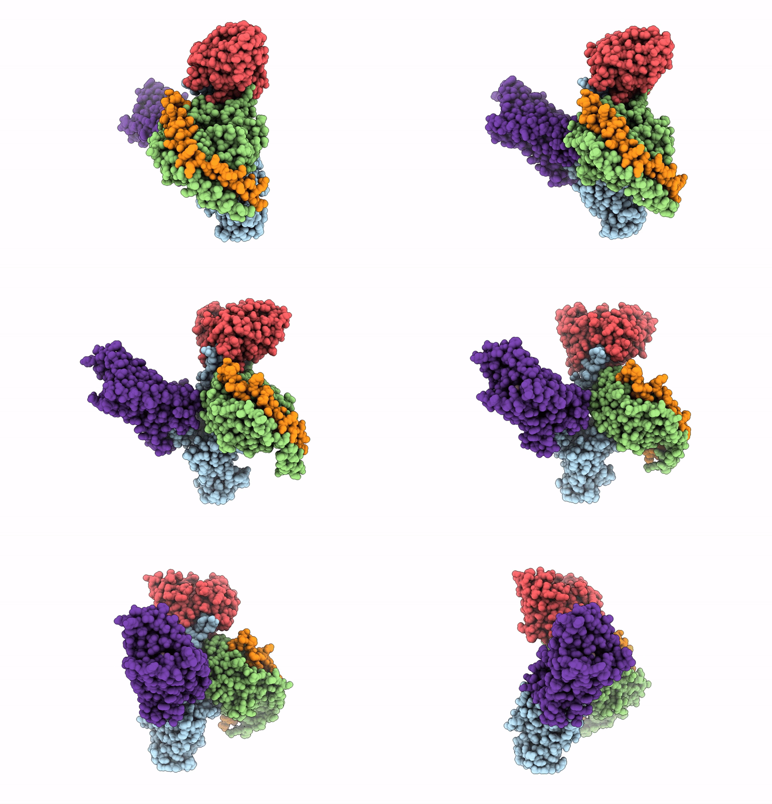
9EBH image
Deposition Date 2024-11-12
Release Date 2025-08-20
Last Version Date 2025-08-20
Entry Detail
PDB ID:
9EBH
Title:
Human adenosine A3 receptor Gi1 complex bound to adenosine
Biological Source:
Source Organism(s):
Homo sapiens (Taxon ID: 9606)
Mus musculus (Taxon ID: 10090)
Expression System(s):
Method Details:
Experimental Method:
Resolution:
3.60 Å
Aggregation State:
PARTICLE
Reconstruction Method:
SINGLE PARTICLE
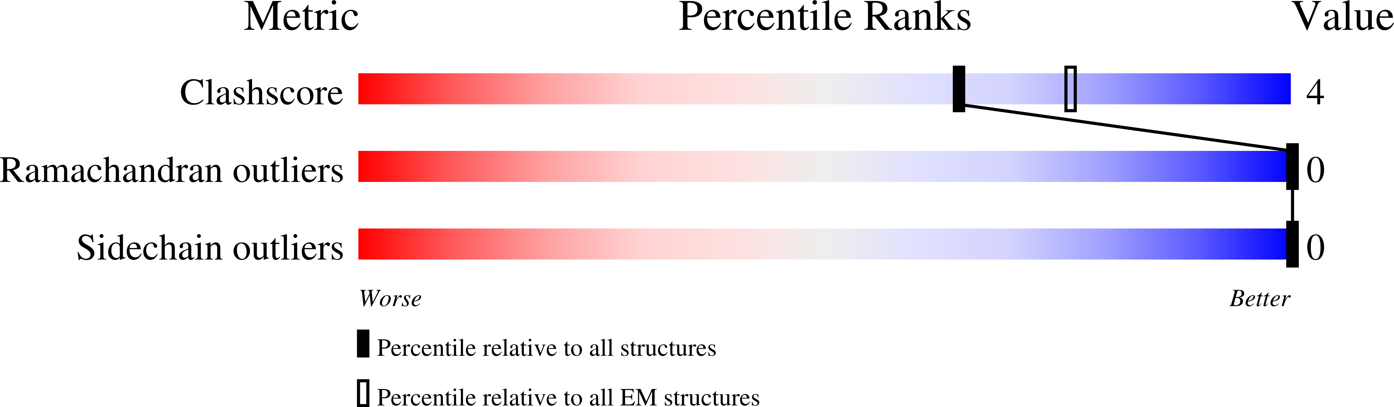
Macromolecular Entities
Structures with similar UniProt ID
Protein Blast
Polymer Type:polypeptide(L)
Molecule:Guanine nucleotide-binding pr
Gene (Uniprot):GNAI1
Chain IDs:A
Chain Length:354
Number of Molecules:1
Biological Source:Homo sapiens
Structures with similar UniProt ID
Protein Blast
Polymer Type:polypeptide(L)
Molecule:Guanine nucleotide-binding pr
Gene (Uniprot):GNB1
Chain IDs:B
Chain Length:349
Number of Molecules:1
Biological Source:Homo sapiens
Protein Blast
Polymer Type:polypeptide(L)
Molecule:Antibody fragment scFv16|Mus
Chain IDs:C (auth: E)
Chain Length:248
Number of Molecules:1
Biological Source:Mus musculus
Structures with similar UniProt ID
Protein Blast
Polymer Type:polypeptide(L)
Molecule:Guanine nucleotide-binding pr
Gene (Uniprot):GNG2
Chain IDs:D (auth: G)
Chain Length:70
Number of Molecules:1
Biological Source:Homo sapiens
Structures with similar UniProt ID
Protein Blast
Polymer Type:polypeptide(L)
Molecule:Adenosine receptor A3
Gene (Uniprot):ADORA3
Chain IDs:E (auth: R)
Chain Length:363
Number of Molecules:1
Biological Source:Homo sapiens
Ligand Molecules
Primary Citation
Molecular basis of ligand binding and receptor activation at the human A 3 adenosine receptor.
Nat Commun 16 7674 7674 (2025)
PMID: 40825947 DOI: 10.1038/s41467-025-62872-x

Abstact

Adenosine receptors (ARs: A1AR, A2AAR, A2BAR, and A3AR) are crucial therapeutic targets; however, developing selective, efficacious drugs for them remains a significant challenge. Here, we present high-resolution cryo-electron microscopy (cryo-EM) structures of the human A3AR in three distinct functional states: bound to the endogenous agonist adenosine, the clinically relevant agonist Piclidenoson, and the covalent antagonist LUF7602. These structures, complemented by mutagenesis and pharmacological studies, reveal an A3AR activation mechanism that involves an extensive hydrogen bond network from the extracellular surface down to the orthosteric binding site. In addition, we identify a cryptic pocket that accommodates the N6-iodobenzyl group of Piclidenoson through a ligand-dependent conformational change of M1745.35. Our comprehensive structural and functional characterisation of A3AR advances our understanding of adenosine receptor pharmacology and establishes a foundation for developing more selective therapeutics for various disorders, including inflammatory diseases, cancer, and glaucoma.

Legend

Protein

Chemical

Disease

Primary Citation of related structures
Feedback Form
Name
Email
Institute
Feedback