

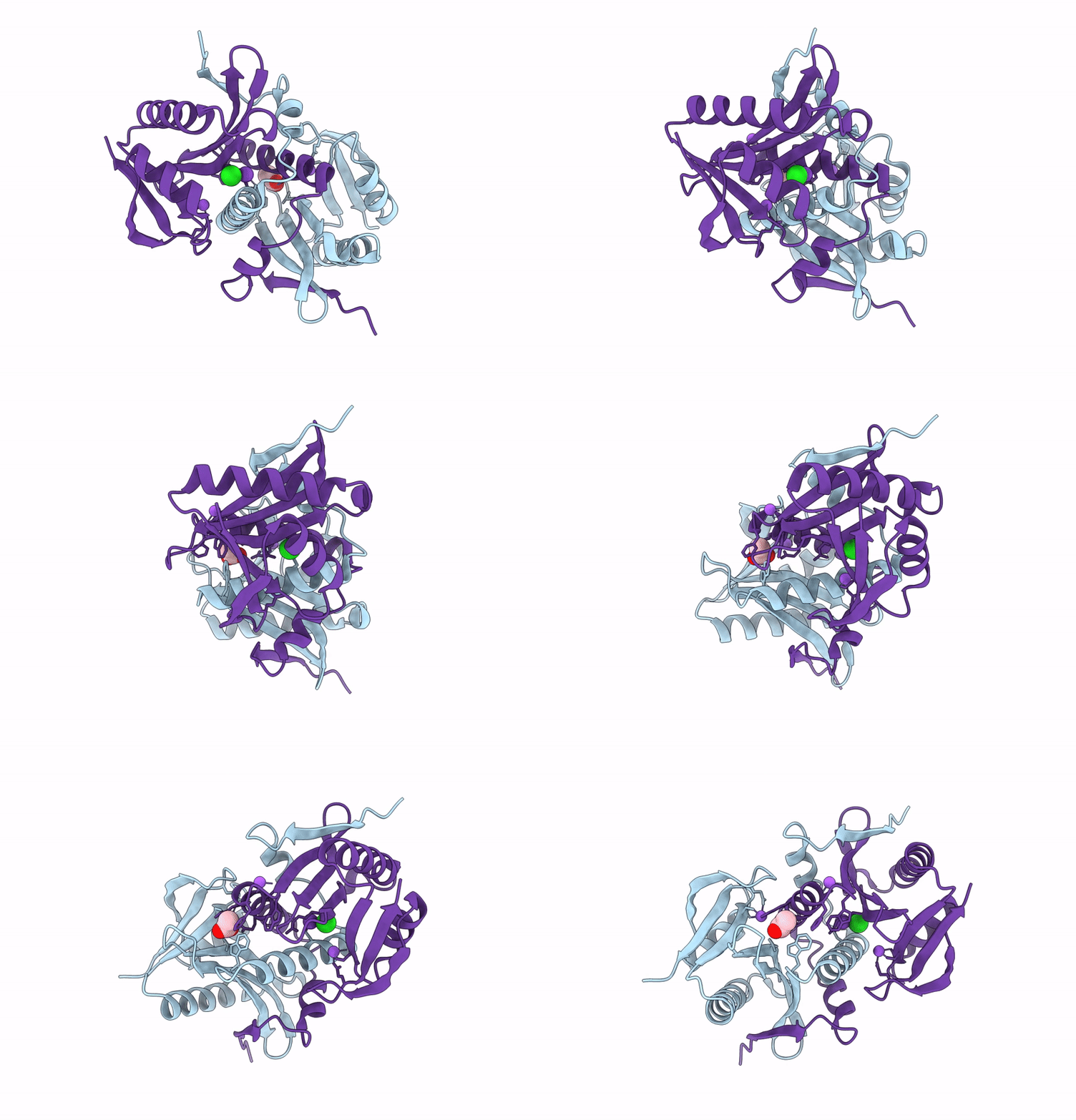
9DSB image
Deposition Date 2024-09-26
Release Date 2025-05-28
Last Version Date 2026-03-25
Entry Detail
PDB ID:
9DSB
Keywords:
Title:
Crystal Structure of Spermin/spermidine N-Acetyltransferase from Enterococcus faecalis V583
Biological Source:
Source Organism(s):
Expression System(s):
Method Details:
Experimental Method:
Resolution:
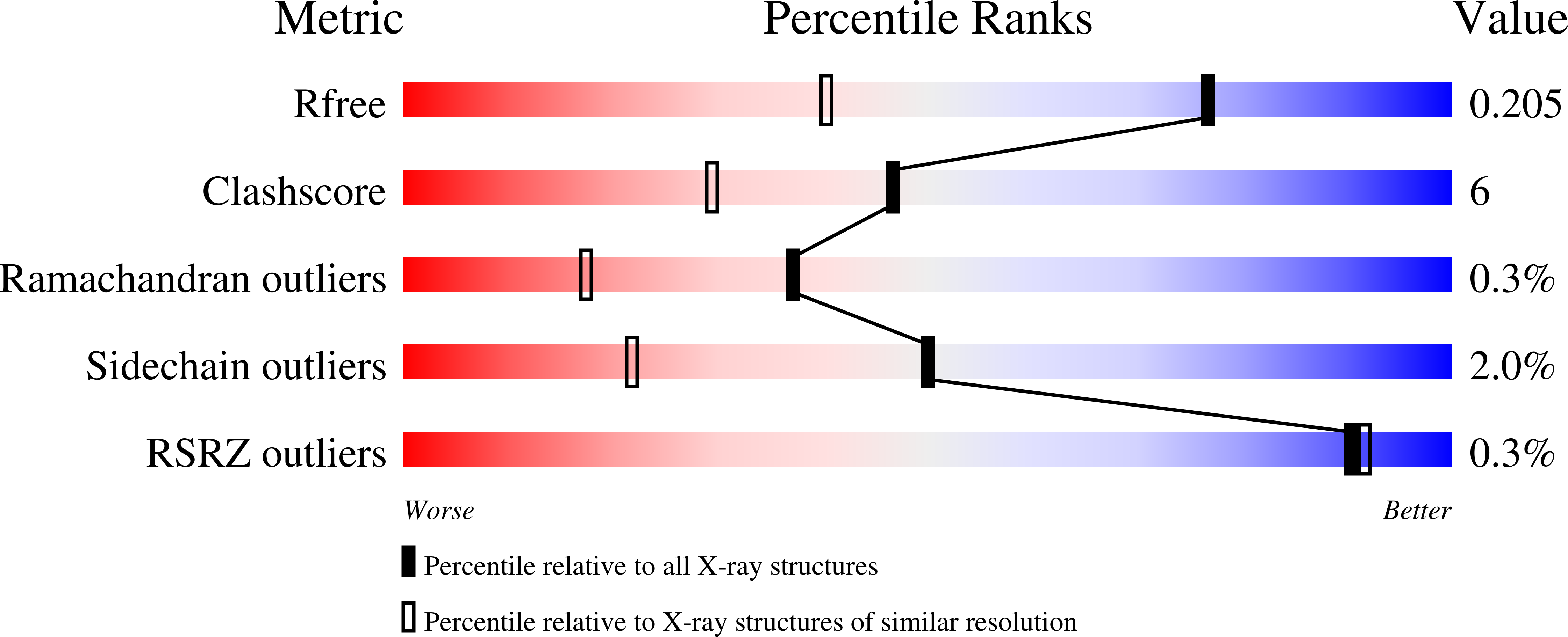
1.50 Å
R-Value Free:
0.20
R-Value Work:
0.16
R-Value Observed:
0.16
Space Group:
P 3
Macromolecular Entities
Structures with similar UniProt ID
Protein Blast
Polymer Type:polypeptide(L)
Molecule:Spermine/spermidine acetyltra
Gene (Uniprot):EF_1086
Chain IDs:A, B
Chain Length:151
Number of Molecules:2
Biological Source:Enterococcus faecalis V583
Primary Citation
N-terminal domain swapping: A new paradigm for spermidine/spermine N-acetyltransferase (SSAT) protein structures?
Biochem.Biophys.Res.Commun. 748 151302 151302 (2025)
PMID: 39823891 DOI: 10.1016/j.bbrc.2025.151302

Abstact

Enterococcus faecalis is a multi-drug-resistant human pathogen that is found in a variety of environments and is challenging to treat. Under stress conditions, some bacteria regulate intracellular polyamine concentrations via polyamine acetyltransferases to reduce their toxicity. The E. faecalis genome encodes two polyamine acetyltransferases: PmvE and BltD. Both of these proteins belong to the Gcn5-related N-acetyltransferase (GNAT) superfamily. It is unclear why there are two enzymes with similar substrate specificities in this organism. To better understand the structure/function relationship of the E. faecalis BltD enzyme, we determined its crystal structure and performed additional assays to explore its oligomeric state and enzymatic activity. The goal was to determine whether there were structural or catalytic differences between this enzyme and other polyamine acetyltransferases that could explain this redundancy and be exploited for future development of targeted inhibitors for this important human pathogen. We found the BltD enzyme was structurally unique due to its N-terminal domain swapped dimer. However, this enzyme adopts a catalytically active monomer rather than dimer in solution. This indicates the crystal structure we obtained may represent a state that forms at high protein and salt concentrations and at low pH used during crystallization. The BltD dimer found in the crystal may represent a unique view of how an inhibitory peptide or molecule could be designed to occupy its active site. Additionally, this structure shows the extensive flexibility of the N-terminal portion of the E. faecalis BltD enzyme.

Legend

Protein

Chemical

Disease

Primary Citation of related structures
Feedback Form
Name
Email
Institute
Feedback