

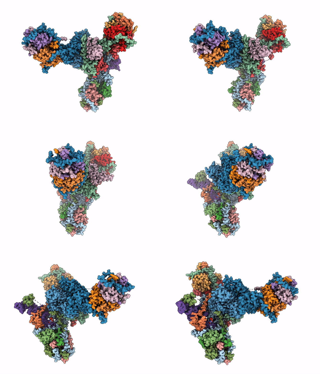
9DEI image
Deposition Date 2024-08-29
Release Date 2026-01-21
Last Version Date 2026-08-05
Entry Detail
PDB ID:
9DEI
Title:
Trypanosoma brucei mitochondrial RNA-editing catalytic complex 1, U-deletion (RECC1)
Biological Source:
Source Organism(s):
Method Details:
Experimental Method:
Resolution:
2.99 Å
Aggregation State:
PARTICLE
Reconstruction Method:
SINGLE PARTICLE
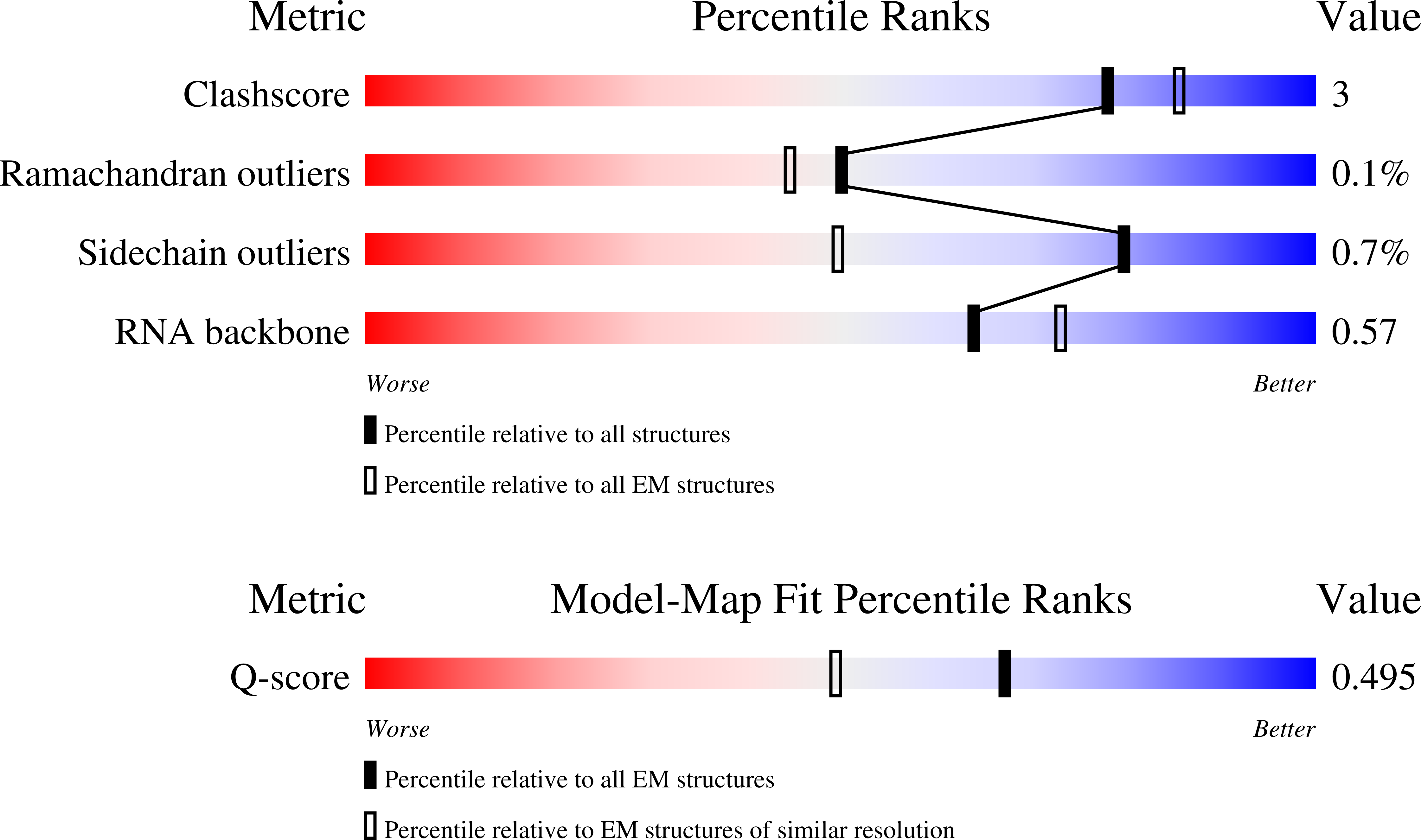
Macromolecular Entities
Polymer Type:polypeptide(L)
Molecule:KREPB5; RNA-editing catalytic
Chain IDs:A
Chain Length:384
Number of Molecules:1
Biological Source:Trypanosoma brucei
Polymer Type:polypeptide(L)
Molecule:KREPB8; RNA editing catalytic
Gene (Uniprot):TbgDal_VIII5690
Chain IDs:B
Chain Length:368
Number of Molecules:1
Biological Source:Trypanosoma brucei
Polymer Type:polypeptide(L)
Molecule:MP90
Gene (Uniprot):TbgDal_I900
Chain IDs:C
Chain Length:818
Number of Molecules:1
Biological Source:Trypanosoma brucei
Polymer Type:polypeptide(L)
Molecule:RNA editing complex protein M
Gene (Uniprot):MP46
Chain IDs:D
Chain Length:414
Number of Molecules:1
Biological Source:Trypanosoma brucei
Polymer Type:polypeptide(L)
Molecule:MP18 RNA editing complex prot
Gene (Uniprot):TbgDal_X6190
Chain IDs:E, H, J, M, P
Chain Length:164
Number of Molecules:5
Biological Source:Trypanosoma brucei
Polymer Type:polypeptide(L)
Molecule:RNA-editing complex protein M
Chain IDs:F, N
Chain Length:393
Number of Molecules:2
Biological Source:Trypanosoma brucei
Polymer Type:polypeptide(L)
Molecule:KREPA2
Gene (Uniprot):KREPA2
Chain IDs:G, K
Chain Length:587
Number of Molecules:2
Biological Source:Trypanosoma brucei
Polymer Type:polypeptide(L)
Molecule:RNA editing complex protein
Gene (Uniprot):TbgDal_X6180
Chain IDs:I
Chain Length:218
Number of Molecules:1
Biological Source:Trypanosoma brucei
Polymer Type:polypeptide(L)
Molecule:KREPA5
Gene (Uniprot):KREPA5
Chain IDs:L
Chain Length:169
Number of Molecules:1
Biological Source:Trypanosoma brucei
Polymer Type:polypeptide(L)
Molecule:RNA-editing complex protein M
Chain IDs:O
Chain Length:762
Number of Molecules:1
Biological Source:Trypanosoma brucei
Polymer Type:polyribonucleotide
Molecule:tRNA-valine (anticodon AAC)
Chain IDs:Q (auth: R)
Chain Length:76
Number of Molecules:1
Biological Source:Trypanosoma brucei
Primary Citation
Structural basis of the RNA-editing cascade in trypanosome mitochondria.
Nature ? ? ? (2026)
PMID: 42486989 DOI: 10.1038/s41586-026-10831-x

Abstact

The molecular mechanism of uridine insertion-and-deletion mRNA editing in trypanosome mitochondria(1-4) has remained unclear because of the highly dynamic nature of the underlying multi-enzyme machinery(5). Here, we define editosomes as supramolecular assemblies formed by the RNA-editing substrate-binding complex (RESC) and either RNA-editing catalytic complex 1 or 2 (RECC1 or RECC2)(6), and present cryo-electron microscopy structures of the approximately 1-MDa RECC1 and RECC2. Resembling dragonflies, with a head, thorax-like core, tail and wings, these ribonucleoproteins mediate the uridine deletion and uridine insertion cascades, respectively. In each RECC, a tetrameric core containing one active and three inactive RNase III domains captures the guide RNA (gRNA)-mRNA duplex, while auxiliary zinc fingers distinguish deletion sites from insertion sites and position the substrate for mRNA cleavage (step I). Three peripheral oligonucleotide-binding-fold heterotetramers are flexibly attached to the core, forming a spatially adaptable reaction chamber. The tail recruits the exonuclease and uridylyltransferase that remove or add uridines (step II), whereas the wings, coordinated by an architectural tRNA, position RNA ligases to seal the edited mRNA (step III). Together, these structures reveal how gRNA-directed substrate recognition, mRNA cleavage, uridine deletion and insertion and ligation are integrated in a single macromolecular machine. This architecture defines the mechanism of information transfer in RNA editing.

Legend

Protein

Chemical

Disease

Primary Citation of related structures
Feedback Form
Name
Email
Institute
Feedback