Deposition Date
2024-08-24
Release Date
2025-08-27
Last Version Date
2026-03-18
Entry Detail
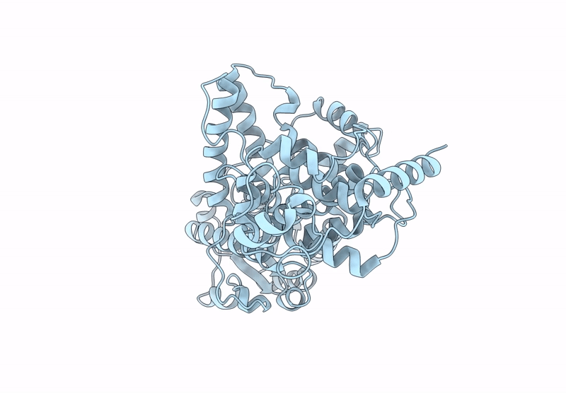
Biological Source:
Source Organism(s):
Erithacus rubecula (Taxon ID: 37610)
Expression System(s):
Method Details:
Experimental Method:
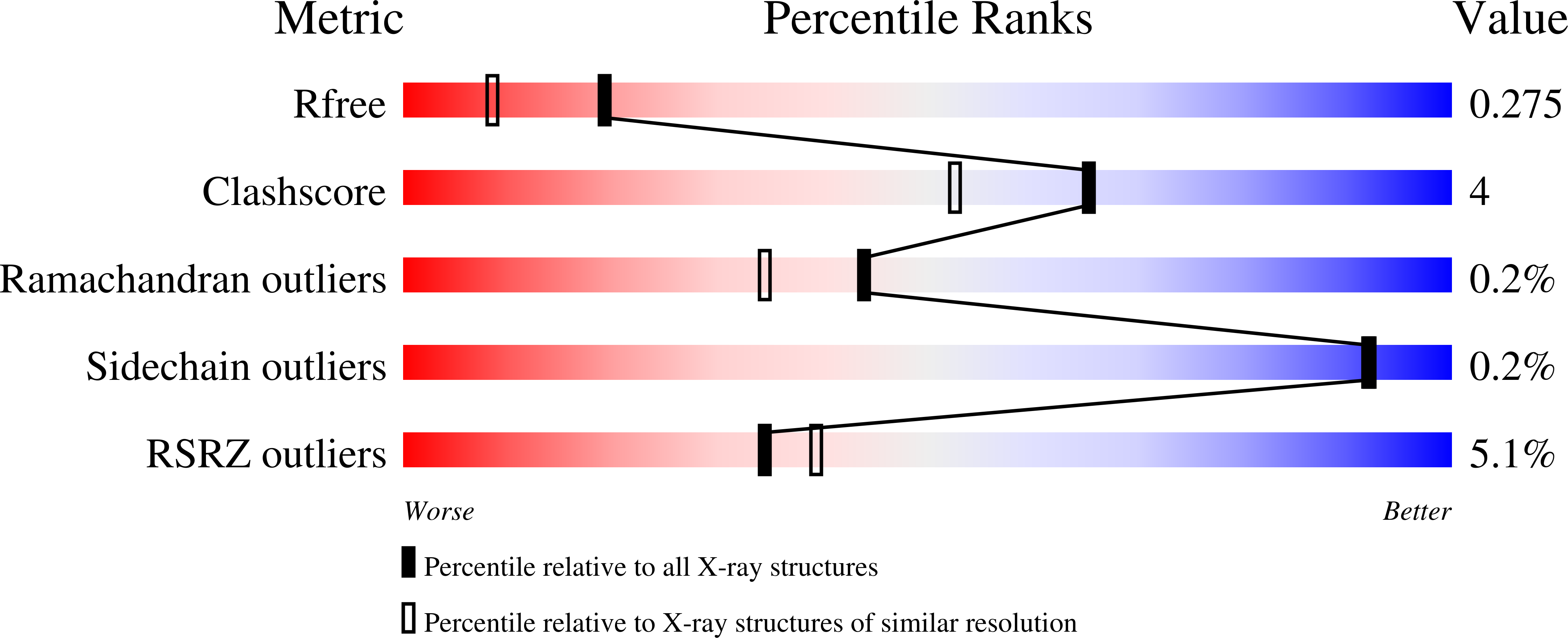
Resolution:
1.91 Å
R-Value Free:
0.27
R-Value Work:
0.20
R-Value Observed:
0.21
Space Group:
P 21 21 21