

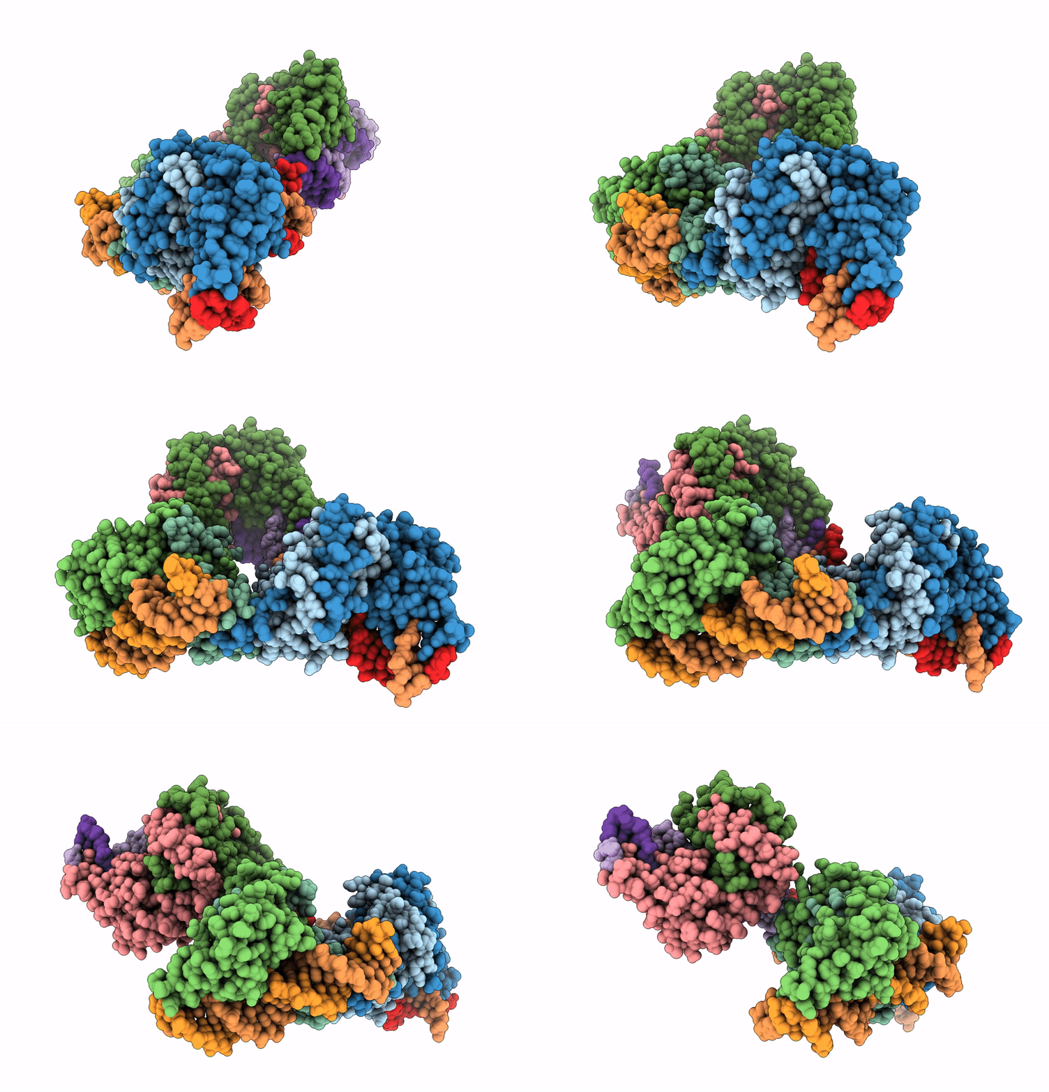
8ZDE image
Deposition Date 2024-05-02
Release Date 2025-11-05
Last Version Date 2026-05-13
Entry Detail
PDB ID:
8ZDE
Keywords:
Title:
Crystal structure of HsmR with DNA bound
Biological Source:
Source Organism(s):
Expression System(s):
Method Details:
Experimental Method:
Resolution:
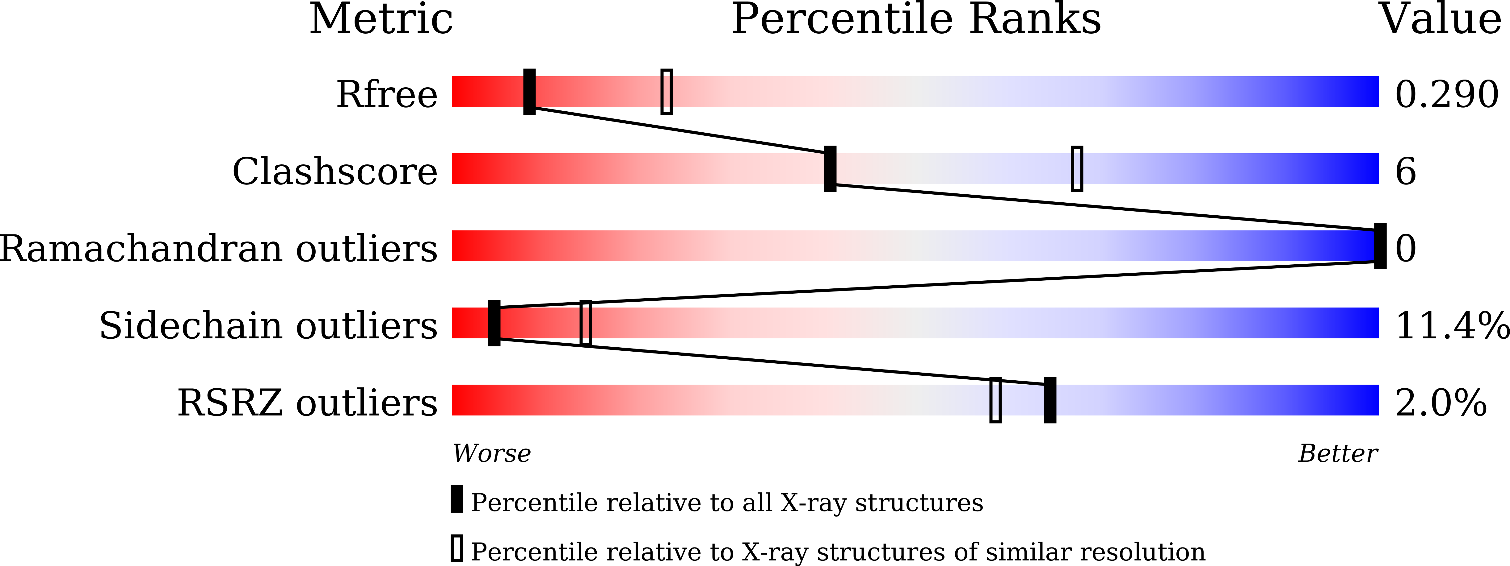
2.91 Å
R-Value Free:
0.27
R-Value Work:
0.21
R-Value Observed:
0.21
Space Group:
C 1 2 1
Macromolecular Entities
Structures with similar UniProt ID
Protein Blast
Polymer Type:polypeptide(L)
Molecule:MarR-family transcriptional r
Gene (Uniprot):CDR20291_0782
Chain IDs:A, B, C, D, E, F
Chain Length:167
Number of Molecules:6
Biological Source:Clostridioides difficile
Polymer Type:polydeoxyribonucleotide
Molecule:DNA (5'-D(*AP*TP*TP*AP*GP*TP*
Chain IDs:G, I, K
Chain Length:22
Number of Molecules:3
Biological Source:Clostridioides difficile
Polymer Type:polydeoxyribonucleotide
Molecule:DNA (5'-D(*AP*AP*TP*GP*GP*TP*
Chain IDs:H, J, L
Chain Length:22
Number of Molecules:3
Biological Source:Clostridioides difficile
Ligand Molecules
Primary Citation
DNA-bound structure of Clostridioides difficile heme-sensing HsmR gives insight on how the unique dimer mode governs DNA specificity in MarR transcriptional regulators.
Nucleic Acids Res. 53 ? ? (2025)
PMID: 41123208 DOI: 10.1093/nar/gkaf1032

Abstact

Clostridioides difficile is a pathogenic bacterium responsible for illnesses ranging from diarrhea to life-threatening colitis and has emerged as a significant public health concern due to its resistance to antibiotics. During its infection, intestinal bleeding causes lysis of the red blood cells releasing heme, a toxic oxidant for the bacterium. To counteract, HsmR of the MarR family transcriptional regulator senses the heme and induces the expression of HsmA to sequester the heme. The structure of HsmR in complex with its cognate pseudo-palindromic DNA illustrates that the lysine and arginine of the winged helix-turn-helix motif undergo conformational changes to accommodate the DNA, and to interact with specific DNA bases. However, conservation of these residues in half of 14 or so C. difficile MarRs within its genome raises a question on how specificity between MarR and DNA is achieved. Comparisons of various C. difficile MarR structures suggest that they probably have acquired DNA selectivity by the slightly different dimeric mode mediated by mutual interaction between the first helices of each HsmR subunit. The unique HsmR dimer mode allows symmetric recognition toward its own cognate DNA, and heme binding would happen in concert with reorientation of these helices in turn affecting DNA binding.

Legend

Protein

Chemical

Disease

Primary Citation of related structures
Feedback Form
Name
Email
Institute
Feedback