

8YFH image
Deposition Date 2024-02-24
Release Date 2025-02-26
Last Version Date 2026-03-11
Entry Detail
PDB ID:
8YFH
Keywords:
Title:
Structure of alpha-1,3-glucanase agn1
Biological Source:
Source Organism(s):
Expression System(s):
Method Details:
Experimental Method:
Resolution:
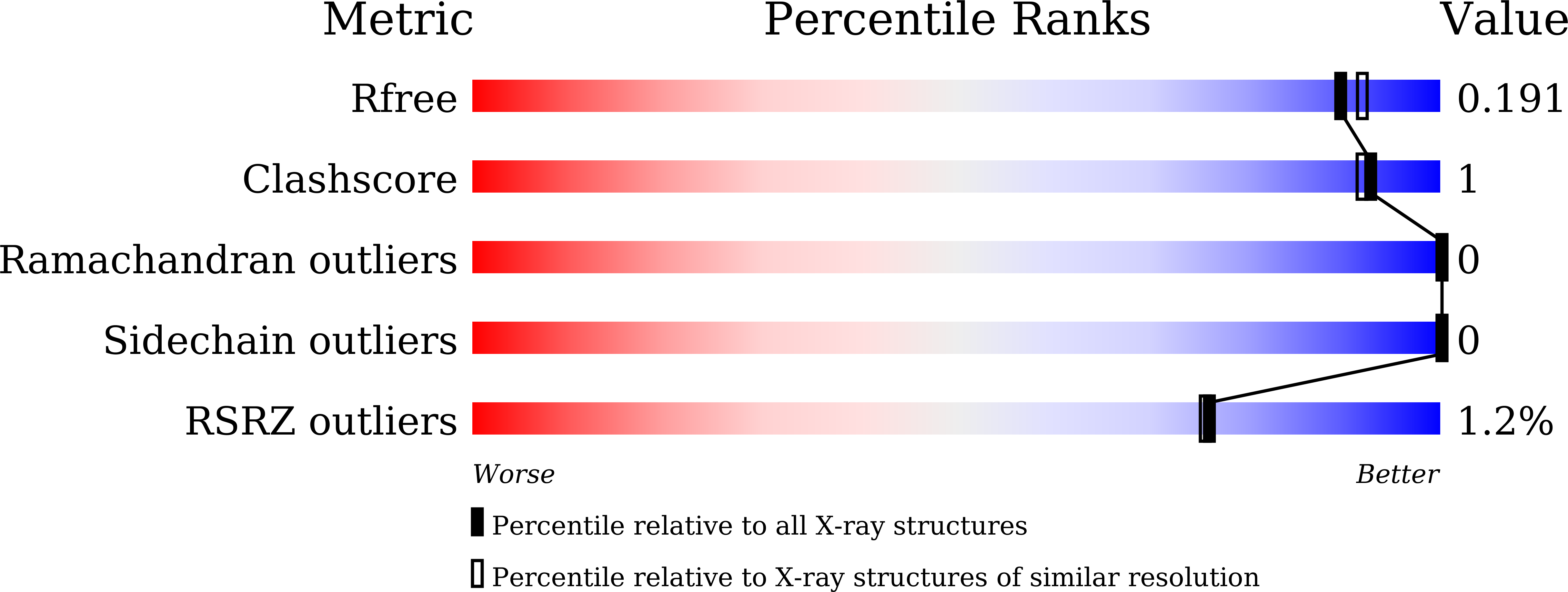
1.80 Å
R-Value Free:
0.19
R-Value Work:
0.16
R-Value Observed:
0.16
Space Group:
P 1 21 1
Macromolecular Entities
Polymer Type:polypeptide(L)
Molecule:Glucan endo-1,3-alpha-glucosi
Gene (Uniprot):agn1
Chain IDs:A, B, C
Chain Length:402
Number of Molecules:3
Biological Source:Schizosaccharomyces pombe DM3650
Primary Citation
Crystal structure of GH71 alpha-1,3-glucanase Agn1p from Schizosaccharomyces pombe: an enzyme regulating cell division in fission yeast.
Biochem.Biophys.Res.Commun. 766 151907 151907 (2025)
PMID: 40306164 DOI: 10.1016/j.bbrc.2025.151907

Abstact

Agn1p is a glycoside hydrolase family 71 alpha-1,3-glucanase from Schizosaccharomyces pombe. It is involved in cell division and releases nigero-pentaose from alpha-1,3-glucan as a primary hydrolysate. In this study, we used x-ray crystallography to determine the molecular structure of Agn1p, achieving a resolution of 1.80 A for its free form and 2.10 A for the substrate complex structure of an inactive mutant. We find that Agn1p comprises eight alpha-helices and sixteen beta-strands, and these combined into a classical (alpha/beta)(8) TIM-barrel core domain and a beta-sandwich accessory domain. The TIM-barrel had a deep cavity in the center. Next, to determine which amino acid residues are involved in the catalytic reaction, we conducted substitution experiments on Asp-69, Asp-237, and Glu-240, three residues located in the cavity, preparing the corresponding substitution mutants D69N, D237A, D237N, E240A and E240Q. We found that the far-UV CD spectra of the five substitution mutants were similar to those of wild-type Agn1p, but all five mutants lost alpha-1,3-glucan hydrolyzing activity. We also obtained the cocrystal of the D237N mutant and nigero-heptaose, and its structure was determined. Specifically, we observed the electron density for the hexamer or pentamer sugar portion of nigero-heptaose. Moreover, the substrates were located in the vicinity of Asp-69, Asp-237, and Glu-240. Overall, these results suggest that Agn1p contains a stable substrate binding site for the hexamer or pentamer sugar structure of nigero-oligosaccharide.

Legend

Protein

Chemical

Disease

Primary Citation of related structures
Feedback Form
Name
Email
Institute
Feedback