

8YEQ image
Deposition Date 2024-02-23
Release Date 2024-09-04
Last Version Date 2024-09-04
Entry Detail
PDB ID:
8YEQ
Title:
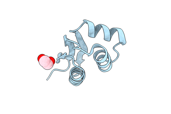
Crystal structure of L7/L12 Ribosomal Protein from Mycobacterium tuberculosis
Biological Source:
Source Organism(s):
Expression System(s):
Method Details:
Experimental Method:
Resolution:
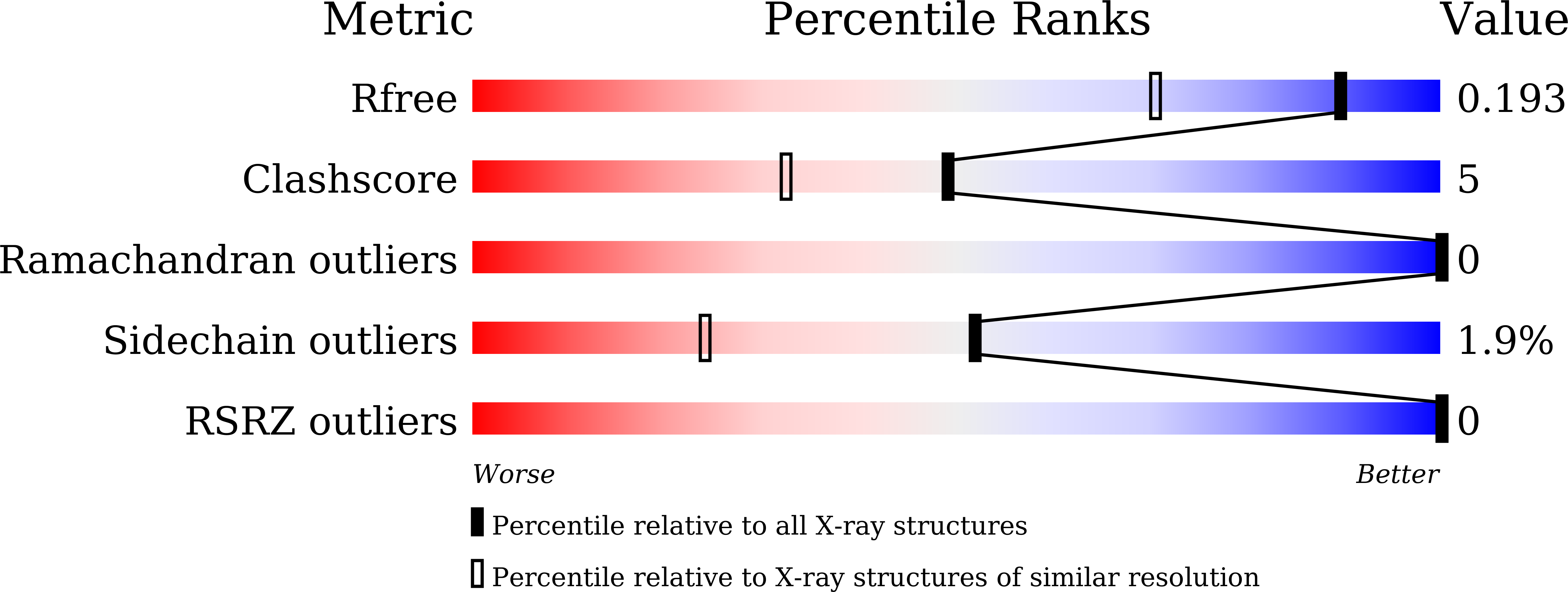
1.50 Å
R-Value Free:
0.19
R-Value Work:
0.16
R-Value Observed:
0.16
Space Group:
P 2 21 21
Macromolecular Entities
Protein Blast
Polymer Type:polypeptide(L)
Molecule:Large ribosomal subunit prote
Gene (Uniprot):rplL
Chain IDs:A
Chain Length:171
Number of Molecules:1
Biological Source:Mycobacterium tuberculosis CDC1551
Ligand Molecules
Primary Citation
Crystal structure, biophysical characterisation, modeling and docking studies of bL12 ribosomal protein from Mycobacterium tuberculosis.
Arch. Biochem. Biophys. 771 110489 110489 (2025)
PMID: 40466900 DOI: 10.1016/j.abb.2025.110489

Abstact

Tuberculosis (TB) is a fatal infectious disease caused by Mycobacterium tuberculosis (Mtb), with high rates of relapse and mortality worldwide. The Mtb stalk protein bL7/bL12 is a component of 50S ribosomal subunit, and plays a crucial role in the translation process during protein synthesis. The bL7 differs from bL12 by the presence of an acetyl group at its N-terminal region. In this study, the bL12 gene from Mtb was cloned into prokaryotic expression vector pET-28a(+), then expressed, purified, characterised and crystallised using the vapour diffusion method. Rod-shaped crystals of bL12 were obtained, which diffracted to 1.5 Å resolution at 100 K, with an Rmerge of 0.025. The bL12 crystallised in the P22121 space group with unit cell dimensions a = 25.86 Å, b = 47.27 Å, c = 61.07 Å, and α = β = γ = 90°. The compact, globular C-terminal domain consists of β1-α1-α2-β2-α3-β3 fold. The bL12 protein was further characterised using various biophysical techniques: CD, SEC, DLS, DSC, DSF and fluorescence spectroscopy. Structural modeling and docking of bL12 protein with its interacting partners in the ribosome were performed using HDOCK and AlphaFold3. The resulting data were analysed to gain insights into its functional role. This structural information on the Mtb bL12 protein enhances our understanding of translational biology and contributes to structure-based drug design efforts targeting tuberculosis.

Legend

Protein

Chemical

Disease

Primary Citation of related structures
Feedback Form
Name
Email
Institute
Feedback