

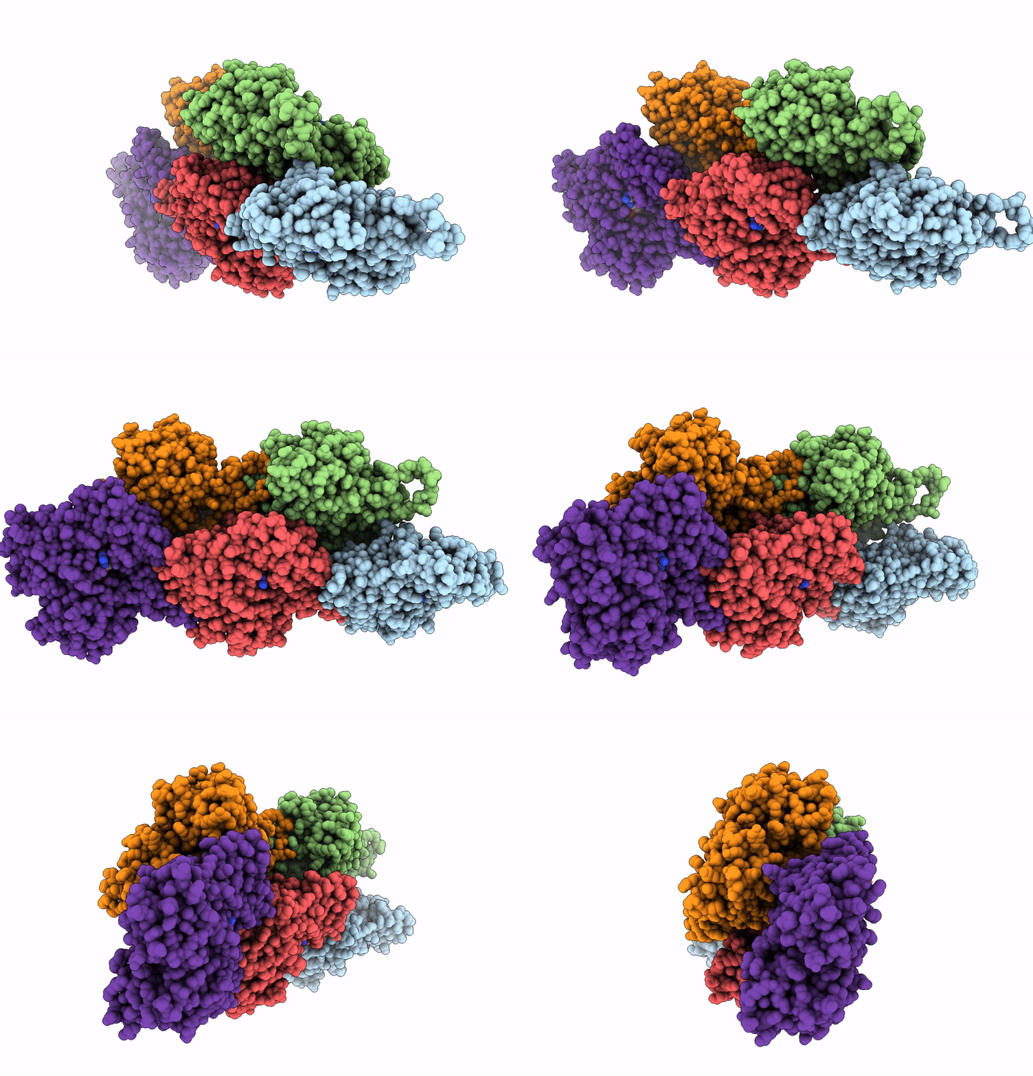
8W36 image
Deposition Date 2024-02-21
Release Date 2025-02-26
Last Version Date 2026-09-16
Entry Detail
PDB ID:
8W36
Title:
rabbit actin in the absence of potassium
Biological Source:
Source Organism(s):
Expression System(s):
Method Details:
Experimental Method:
Resolution:
2.27 Å
Aggregation State:
FILAMENT
Reconstruction Method:
HELICAL
Macromolecular Entities
Polymer Type:polypeptide(L)
Molecule:Actin, alpha skeletal muscle
Gene (Uniprot):ACTA1
Chain IDs:A (auth: D), B (auth: C), C (auth: A), D (auth: B), E
Chain Length:371
Number of Molecules:5
Biological Source:Oryctolagus cuniculus
Modified Residue
Compound ID Chain ID Parent Comp ID Details 2D Image
HIC A, B, C, D, E HIS modified residue
Primary Citation
High-resolution yeast actin structures indicate the molecular mechanism of actin filament stiffening by cations.
Commun Chem 7 164 164 (2024)
PMID: 39079963 DOI: 10.1038/s42004-024-01243-x

Abstact

Actin filament assembly and the regulation of its mechanical properties are fundamental processes essential for eukaryotic cell function. Residue E167 in vertebrate actins forms an inter-subunit salt bridge with residue K61 of the adjacent subunit. Saccharomyces cerevisiae actin filaments are more flexible than vertebrate filaments and have an alanine at this position (A167). Substitution of this alanine for a glutamic acid (A167E) confers Saccharomyces cerevisiae actin filaments with salt-dependent stiffness similar to vertebrate actins. We developed an optimized cryogenic electron microscopy workflow refining sample preparation and vitrification to obtain near-atomic resolution structures of wild-type and A167E mutant Saccharomyces cerevisiae actin filaments. The difference between these structures allowed us to pinpoint the potential binding site of a filament-associated cation that controls the stiffness of the filaments in vertebrate and A167E Saccharomyces cerevisiae actins. Through an analysis of previously published high-resolution reconstructions of vertebrate actin filaments, along with a newly determined high-resolution vertebrate actin structure in the absence of potassium, we identified a unique peak near residue 167 consistent with the binding of a magnesium ion. Our findings show how magnesium can contribute to filament stiffening by directly bridging actin subunits and allosterically affecting the orientation of the DNase-I binding loop of actin, which plays a regulatory role in modulating actin filament stiffness and interactions with regulatory proteins.

Legend

Protein

Chemical

Disease

Primary Citation of related structures
Feedback Form
Name
Email
Institute
Feedback