

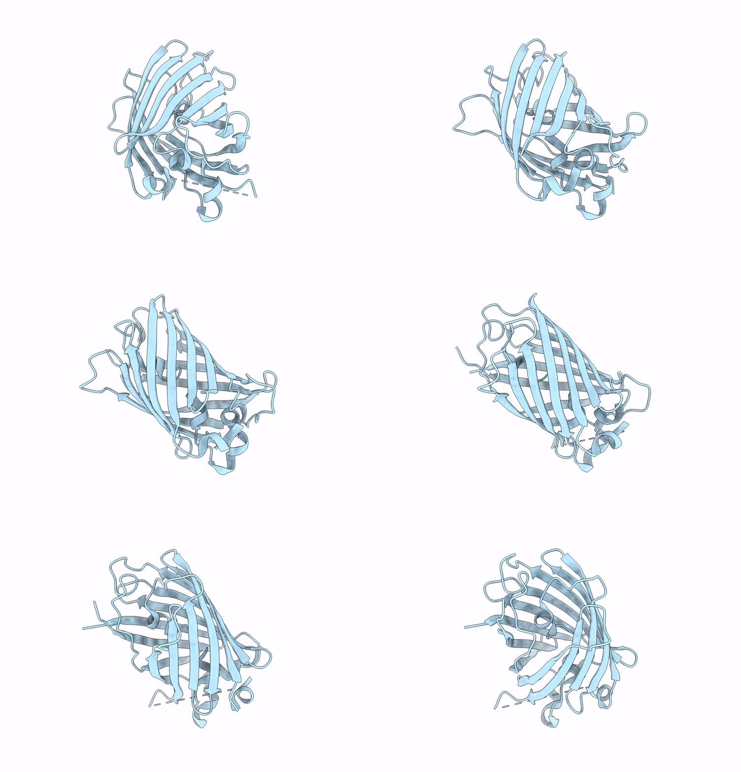
8UQQ image
Deposition Date 2023-10-24
Release Date 2024-10-30
Last Version Date 2026-05-13
Entry Detail
PDB ID:
8UQQ
Title:
Structure of mCLIFY: a circularly permuted yellow fluorescent protein
Biological Source:
Source Organism(s):
Expression System(s):
Method Details:
Experimental Method:
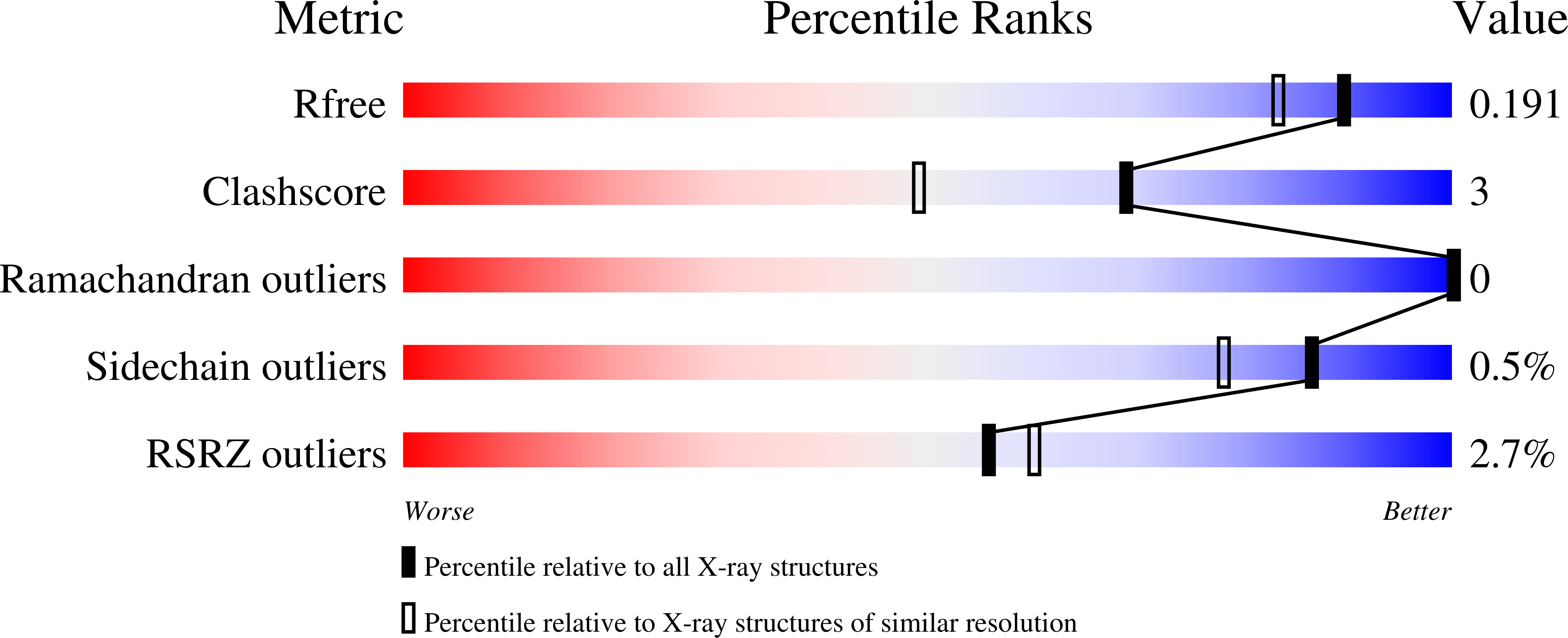
Resolution:
1.57 Å
R-Value Free:
0.19
R-Value Work:
0.17
R-Value Observed:
0.17
Space Group:
P 2 21 21
Macromolecular Entities
Polymer Type:polypeptide(L)
Molecule:mCLIFY
Chain IDs:A
Chain Length:253
Number of Molecules:1
Biological Source:Aequorea victoria
Primary Citation
Characterization and structural basis for the brightness of mCLIFY: A novel monomeric and circularly permuted bright yellow fluorescent protein.
Biophys.J. 124 2133 2149 (2025)
PMID: 40405461 DOI: 10.1016/j.bpj.2025.05.012

Abstact

Ongoing improvements of genetically encoded fluorescent proteins have enhanced cellular localization studies and performance of biosensors, such as environmentally or mechanically sensitive fluorescence resonance energy transfer pairs, in cell biological and biophysical research. The brightest yellow fluorescent protein, widely used in these studies is YPet, derived from the jellyfish Aequorea victoria via the GFP derivative Venus. YPet dimerizes at concentrations used in cellular studies (K(D)(1-2) = 3.4 muM) which impacts quantitative interpretation of emission intensity, rotational freedom, energy transfer, and lifetime. Although YPet is nearly 30% brighter than Venus, no atomic structures of YPet have been reported to ascertain the structural differences leading to the higher brightness, possibly due to the tendency to dimerize or oligomerize. Here, we report properties of a new YPet derivative, mCLIFY, a monomeric, bright, yellow, and long-lived fluorescent protein created by circular permutation of YPet and substitution of the amino acid residues thought to mediate dimerization. mCLIFY retains the advantageous photophysical properties of YPet but does not dimerize at least up to 40 muM concentration. We determined the atomic structure of mCLIFY at 1.57-A resolution. Extensive characterization of the photophysical and structural properties of YPet and mCLIFY allowed us to elucidate the bases of their long lifetimes, enhanced brightness, and the difference in propensity to dimerize.

Legend

Protein

Chemical

Disease

Primary Citation of related structures
Feedback Form
Name
Email
Institute
Feedback