

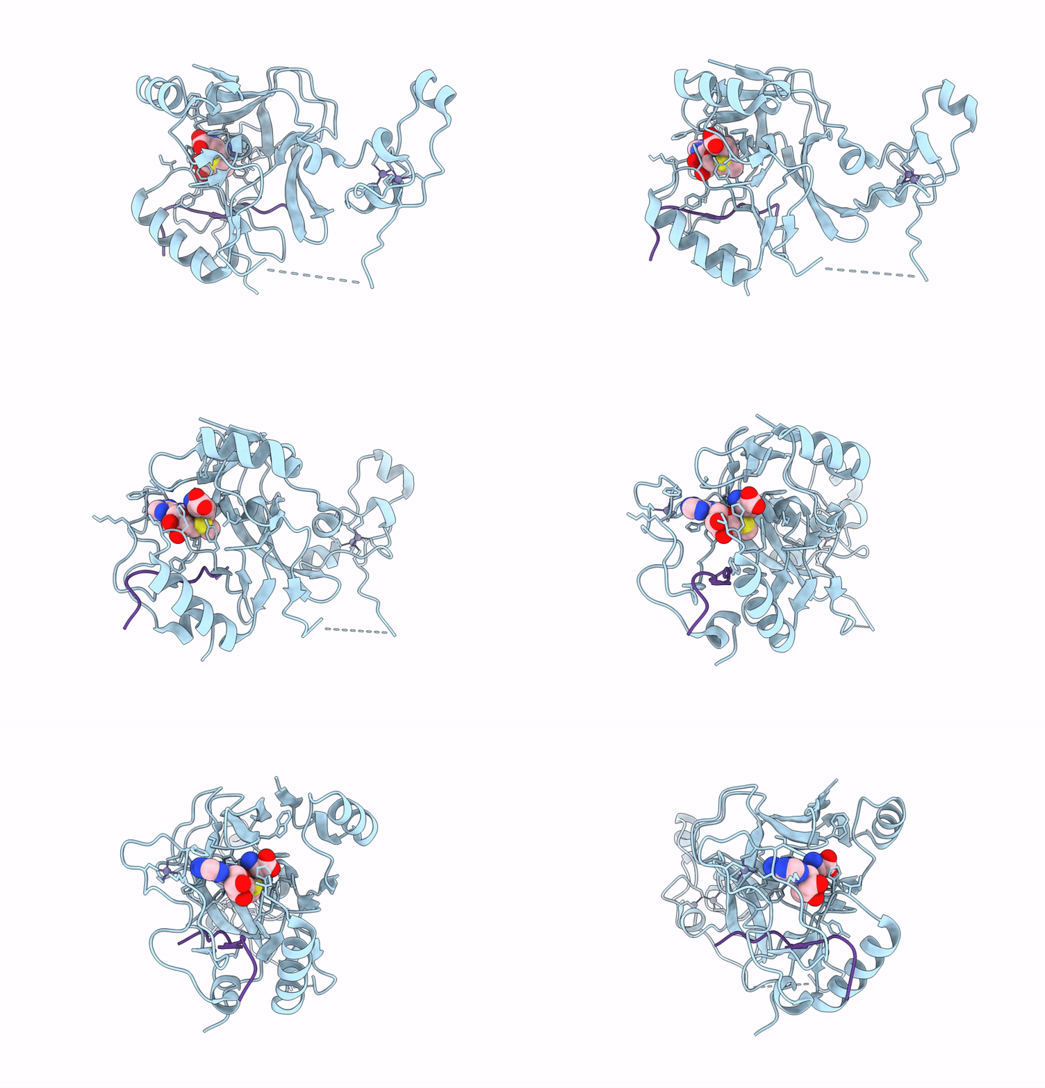
8RZU image
Deposition Date 2024-02-13
Release Date 2025-03-05
Last Version Date 2026-03-18
Entry Detail
PDB ID:
8RZU
Keywords:
Title:
Structure of human SETD2 L1609P mutant in complex with SAM and H3K36M peptide
Biological Source:
Source Organism(s):
Homo sapiens (Taxon ID: 9606)
Expression System(s):
Method Details:
Experimental Method:
Resolution:
2.19 Å
R-Value Free:
0.26
R-Value Work:
0.22
R-Value Observed:
0.22
Space Group:
P 21 21 21

Macromolecular Entities
Structures with similar UniProt ID
Protein Blast
Polymer Type:polypeptide(L)
Molecule:Histone-lysine N-methyltransf
Gene (Uniprot):SETD2
Mutagens:L1609P
Chain IDs:A
Chain Length:295
Number of Molecules:1
Biological Source:Homo sapiens
Structures with similar UniProt ID
Protein Blast
Polymer Type:polypeptide(L)
Molecule:Histone H3
Chain IDs:B
Chain Length:14
Number of Molecules:1
Biological Source:Homo sapiens
Primary Citation
The SETD2 L1609P mutation found in leukemia disrupts methyltransferase activity and reduces histone H3K36 trimethylation.
J. Biol. Chem. 302 111259 111259 (2026)
PMID: 41654133 DOI: 10.1016/j.jbc.2026.111259

Abstact

SET-domain containing protein 2 (SETD2) is the primary methyltransferase responsible for generating H3K36me3, an epigenetic mark that is essential for transcriptional regulation and chromatin integrity. SETD2 mutations are frequently observed in various cancers and tend to cluster within its catalytic SET domain. Despite the clinical relevance of SETD2 missense mutations in cancer, their biochemical and structural consequences remain insufficiently characterized. Here, we present the enzymatic and structural characterization of the SETD2 L1609P mutant enzyme identified in leukemia. The L1609 residue is located in the SET domain within a conserved hydrophobic pocket that is involved in substrate H3K36 recognition. Interestingly, site-directed mutagenesis of residues within this hydrophobic pocket leads to SETD2 enzyme variants with either decreased or increased H3K36me3 methyltransferase activity, suggesting that cancer mutations affecting the L1609 residue could result in a loss- or gain-of-function enzyme variant. Using molecular and cellular approaches, we show that the SETD2 L1609P mutant exhibits reduced H3K36 methyltransferase activity, decreased protein stability, and poor cellular expression. Consistently, the crystal structure of the SETD2 L1609P in complex with a H3K36M peptide shows remodeling of the active site. These findings support the pivotal role of SETD2 inactivation and subsequent disruption of H3K36me3 deposition in oncogenesis, particularly in hematologic malignancies. Our study provides the first mechanistic and three-dimensional protein structure information on how SETD2-associated cancer mutations can lead to altered H3K36 methyltransferase activity.

Legend

Protein

Chemical

Disease

Primary Citation of related structures
Feedback Form
Name
Email
Institute
Feedback