

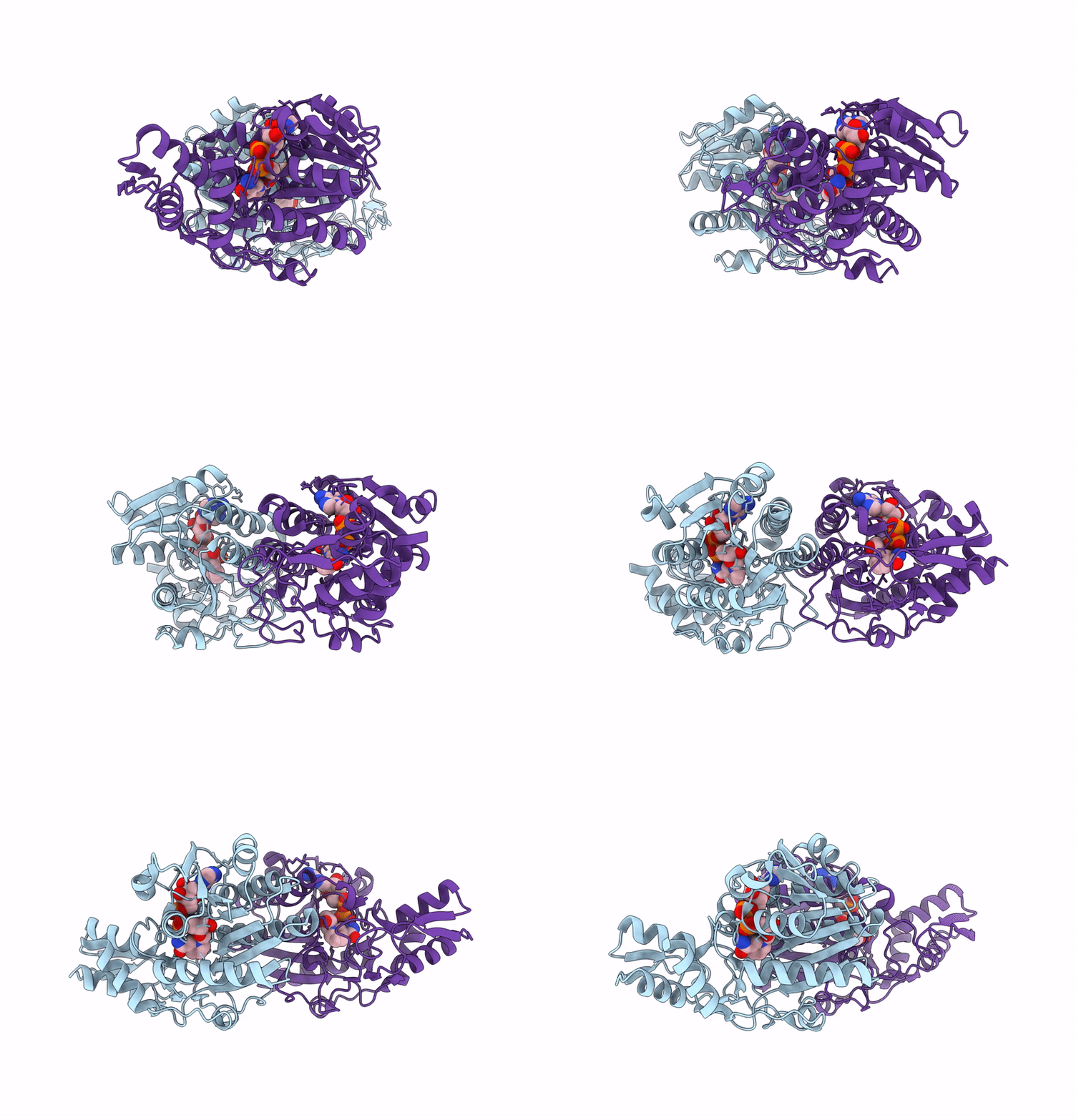
8RDG image
Deposition Date 2023-12-08
Release Date 2024-12-18
Last Version Date 2026-07-01
Entry Detail
PDB ID:
8RDG
Keywords:
Title:
Crystal structure of UDP-galactose 4-epimerase from Pyrococcus horikoshii with bound NAD
Biological Source:
Source Organism(s):
Expression System(s):
Method Details:
Experimental Method:
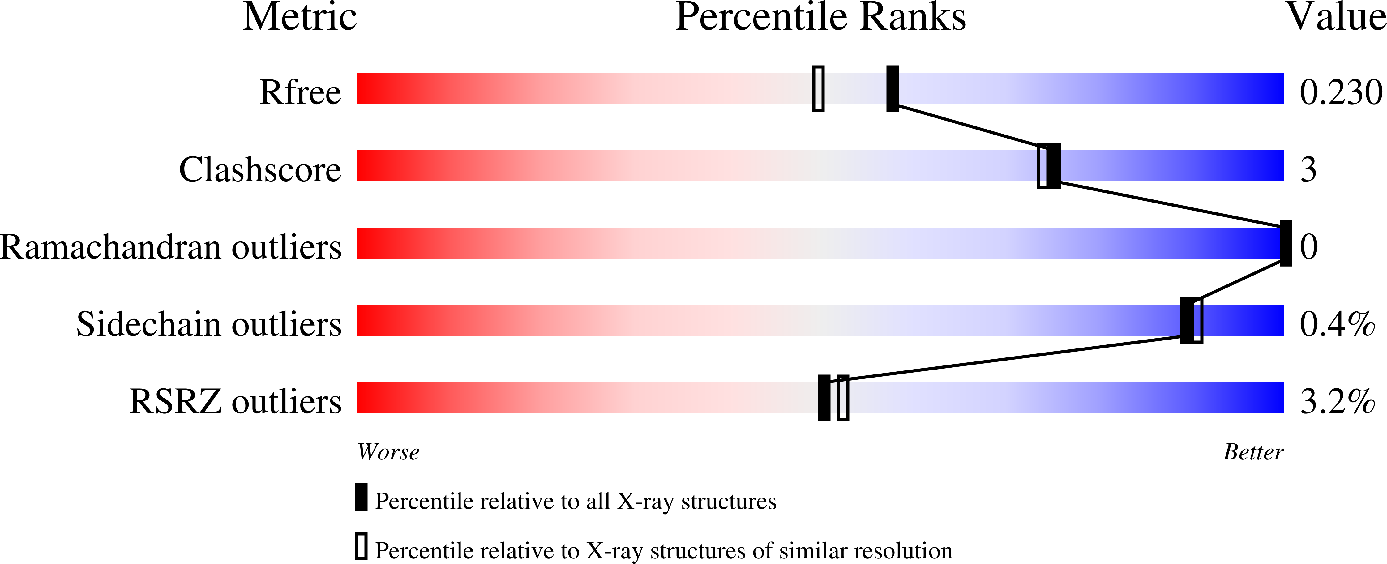
Resolution:
1.90 Å
R-Value Free:
0.23
R-Value Work:
0.19
R-Value Observed:
0.19
Space Group:
P 21 21 21
Macromolecular Entities
Structures with similar UniProt ID
Protein Blast
Polymer Type:polypeptide(L)
Molecule:SDR family NAD(P)-dependent o
Chain IDs:A, B
Chain Length:326
Number of Molecules:2
Biological Source:Pyrococcus horikoshii
Ligand Molecules
Primary Citation
Protein flexibility drives sugar rotation and high substrate promiscuity in a GDP-sugar 4-epimerase.
Comput Struct Biotechnol J 27 2375 2385 (2025)
PMID: 40529184 DOI: 10.1016/j.csbj.2025.05.037

Abstact

UDP-galactose 4-epimerases (Gal4Es) catalyze the inversion of the 4-hydroxyl configuration of a sugar moiety from an NDP-sugar through a three-step process: oxidation, rotation and reduction. Despite extensive biochemical and structural studies, the role of protein dynamics on substrate specificity remains poorly understood. The recently identified subgroup of GDP-sugar 4-epimerases, notable for its exceptional substrate promiscuity, provides an intriguing model to investigate the role of dynamics in the Gal4E catalytic mechanism and the unique promiscuity of the subgroup. In this study, we used a multidisciplinary approach to examine the dynamic-function relationships in the Pyrococcus horikoshii representative (PhGal4E_1). First, we determined several crystal structures (WT: 1.9-2.4 A and Y145F: 3.1 A), providing structural insights of the PhGal4E_1 structure bound to GDP-L-fucose in a catalytic conformation. To further explore the enzyme's promiscuity, in silico docking studies were conducted with three substrates, namely GDP-L-Fuc, GDP-Glc and UDP-Glc. Molecular dynamics simulations identified a dynamic hydrogen bond network surrounding the sugar moiety and phosphate groups, revealing four key residues: P80, H182, R83 and N174. These residues interact with either the substrate's sugar moiety (H182 and P80 with C2-OH and C3-OH, resp.) or diphosphate backbone (N174 and R83 with beta-/alpha- and alpha-phosphate, resp.), which facilitates sugar ring positioning. Protein flexibility then initiates disruption of the hydrogen bonds enabling the required rotation of the intermediate. Site directed mutagenesis of these residues was performed to disrupt the interaction network followed by enzyme activity assays on the three substrates, validating their critical role in the epimerization reaction. These results highlight the pivotal role of protein flexibility in PhGal4E_1 promiscuity and establish a framework for dynamic studies across other Gal4E representatives.

Legend

Protein

Chemical

Disease

Primary Citation of related structures
Feedback Form
Name
Email
Institute
Feedback