

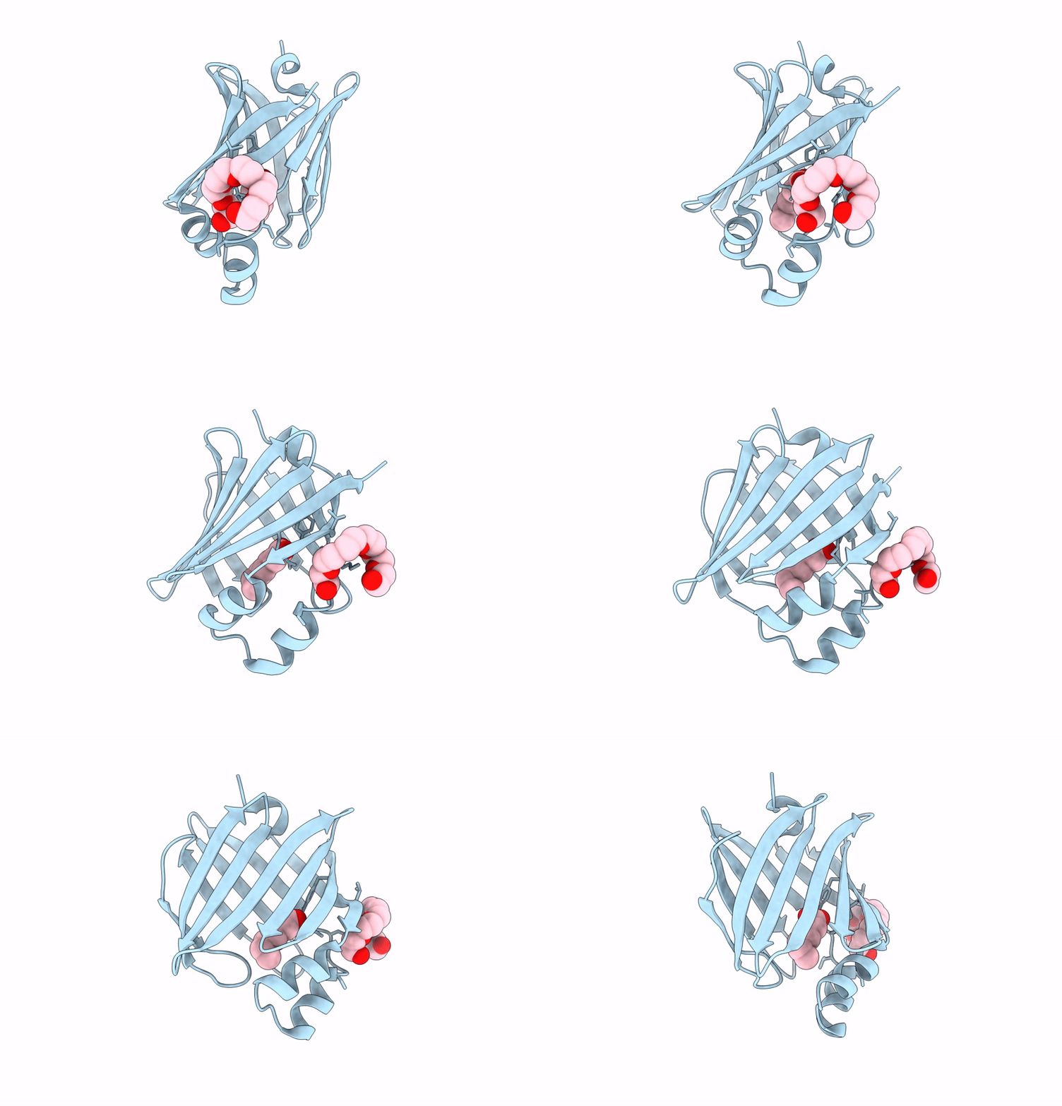
7WPG image
Deposition Date 2022-01-23
Release Date 2023-01-25
Last Version Date 2026-06-24
Entry Detail
PDB ID:
7WPG
Title:
The 0.90 angstrom X-ray structure of the human heart fatty acid-binding protein complexed with heptanoic acid
Biological Source:
Source Organism(s):
Homo sapiens (Taxon ID: 9606)
Expression System(s):
Method Details:
Experimental Method:
Resolution:
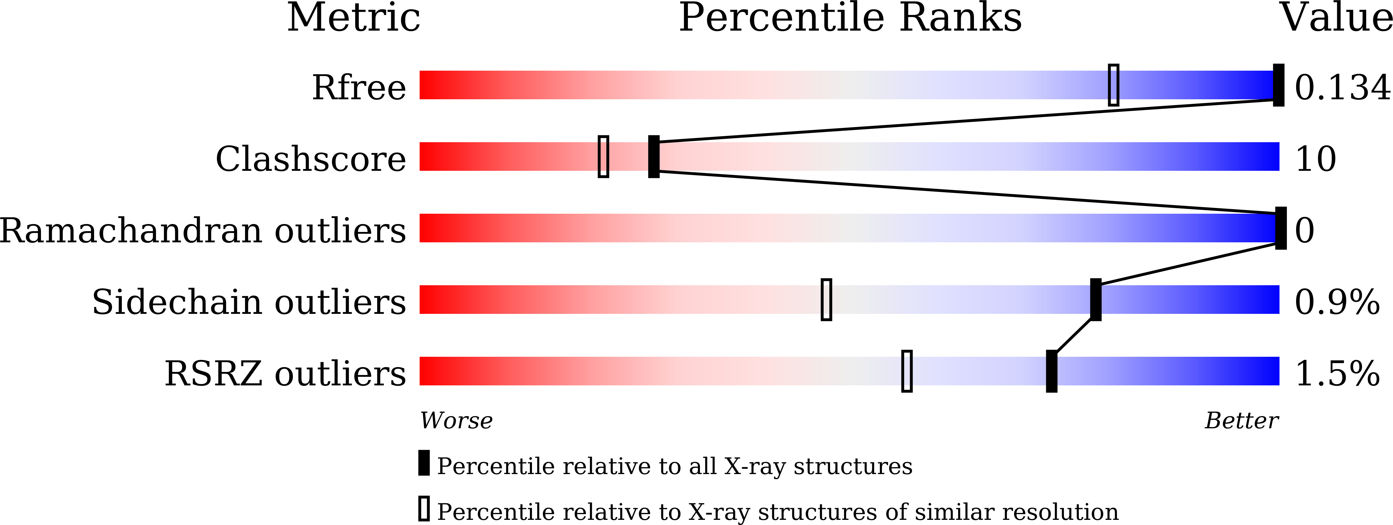
0.90 Å
R-Value Free:
0.12
R-Value Work:
0.11
R-Value Observed:
0.11
Space Group:
P 21 21 21
Macromolecular Entities
Polymer Type:polypeptide(L)
Molecule:Fatty acid-binding protein, h
Gene (Uniprot):FABP3
Chain IDs:A
Chain Length:133
Number of Molecules:1
Biological Source:Homo sapiens
Primary Citation
Intermolecular interactions of perfluoroalkyl acids with human heart-type fatty acid-binding protein.
Int.J.Biol.Macromol. 369 152710 152710 (2026)
PMID: 42190779 DOI: 10.1016/j.ijbiomac.2026.152710

Abstact

PFAS are widely employed in a broad range of applications, spanning from consumer products, such as non-stick cookware, to industrial processes including semiconductor manufacturing. However, PFAS can accumulate in the human body, and certain compounds have been reported to exhibit carcinogenic potential. Perfluoroalkyl acids (PFAAs), a subclass of PFAS, have been shown to bioaccumulate via interactions with fatty acid-binding proteins (FABPs), although the molecular basis for their recognition remains incompletely elucidated. In this study, fluorescence displacement assays revealed that two perfluoroalkyl acids (PFAAs) showed lower apparent IC(5)(0) values for human FABP3 than their corresponding physiological ligands, medium-chain fatty acids (MCFAs). We also determined the ultra-high resolution crystal structures of FABP3 in complex with PFAAs and with MCFAs, thereby providing a molecular basis for PFAAs recognition by FABP3. Structural comparisons demonstrated that PFAAs adopt conformations resembling MCFAs but show distinct solvent-coupled features, including close O...F contacts with ordered water molecules in the binding pocket. Our findings suggest that FABP3 recognizes PFAAs through a mechanism partially shared with fatty acids, but not fully explained by hydrophobic effects alone, with possible additional contributions from dipole-interactive effects. This work provides structural insight into PFAS recognition and suggests a molecular basis by which PFAS could interfere with fatty acid binding to FABPs.

Legend

Protein

Chemical

Disease

Primary Citation of related structures
Feedback Form
Name
Email
Institute
Feedback