Deposition Date
2022-02-04
Release Date
2023-02-15
Last Version Date
2026-05-27
Entry Detail
PDB ID:
7R2D
Keywords:
Title:
Crystal structure of TaCel5A E133A variant in complex with cellopentaose
Biological Source:
Source Organism(s):
Thermoascus aurantiacus ATCC 26904 (Taxon ID: 1133049)
Expression System(s):
Method Details:
Experimental Method:
Resolution:
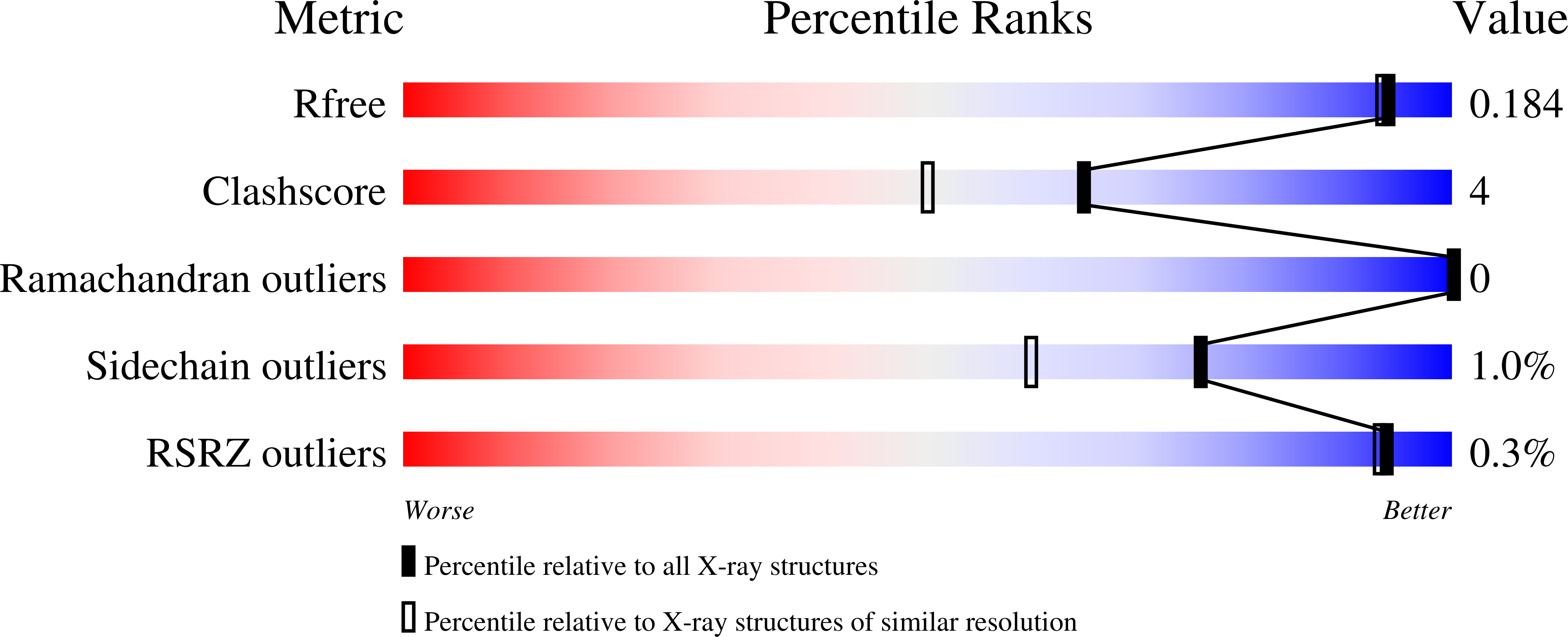
1.61 Å
R-Value Free:
0.18
R-Value Work:
0.15
R-Value Observed:
0.15
Space Group:
P 21 21 21