7JJQ image
Deposition Date 2020-07-27
Release Date 2020-11-11
Last Version Date 2023-10-18
Entry Detail
PDB ID:
7JJQ
Title:
Human Hemoglobin in Complex with Nitrosoamphetamine
Biological Source:
Source Organism(s):
Homo sapiens (Taxon ID: 9606)
Method Details:
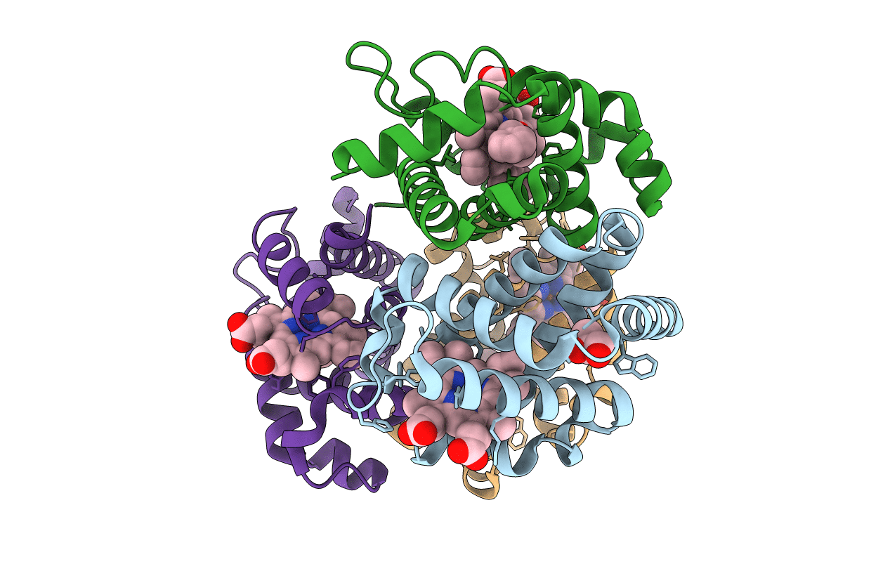
Experimental Method:
Resolution:
2.15 Å
R-Value Free:
0.28
R-Value Work:
0.21
R-Value Observed:
0.22
Space Group:
P 41
Macromolecular Entities
Structures with similar UniProt ID
Protein Blast
Polymer Type:polypeptide(L)
Molecule:Hemoglobin subunit alpha
Gene (Uniprot):HBA1, HBA2
Chain IDs:A, C
Chain Length:141
Number of Molecules:2
Biological Source:Homo sapiens
Structures with similar UniProt ID
Protein Blast
Polymer Type:polypeptide(L)
Molecule:Hemoglobin subunit beta
Gene (Uniprot):HBB
Chain IDs:B, D
Chain Length:146
Number of Molecules:2
Biological Source:Homo sapiens
Primary Citation
The nitrosoamphetamine metabolite is accommodated in the active site of human hemoglobin: Spectroscopy and crystal structure.
J. Inorg. Biochem. 213 111262 111262 (2020)
PMID: 33049600 DOI: 10.1016/j.jinorgbio.2020.111262

Abstact

Amphetamine-based (Amph) drugs are metabolized in humans to their hydroxylamine (AmphNHOH) and nitroso (AmphNO) derivatives. The latter metabolites are known to bind to the Fe centers of cytochrome P450 and other heme enzymes to inhibit their activities. Although these AmphNHOH/AmphNO metabolites are present in vivo, their interactions with the blood protein hemoglobin (Hb) and the muscle protein (Mb) have been largely discounted due to a perception that the relatively small heme active sites of Hb and Mb will not be able to accommodate the large AmphNO group. We report the 2.15 Å resolution X-ray crystal structure of the AmphNO adduct of adult human hemoglobin as the Hb [α-FeIII(H2O)][β-FeII(AmphNO)] derivative. We show that the binding of AmphNO to the β subunit is enabled by an E helix movement and stabilization of ligand binding by H-bonding with the distal His63 residue. We also observe an AmphNHOH group in the Xe2 pocket in close proximity to the α heme site in this derivative. Additionally, UV-vis spectroscopy was used to characterize this and related wt and mutant Mb adducts. Importantly, our X-ray crystal structure of this Hb-nitrosoamphetamine complex represents the first crystal structure of a wild-type heme protein adduct of any amphetamine metabolite. Our results provide a framework for further studies of AmphNHOH/AmphNO interactions with Hb and Mb as viable processes that potentially contribute to the overall biological inorganic chemistry of amphetamine drugs.

Legend

Protein

Chemical

Disease

Primary Citation of related structures
Feedback Form
Name
Email
Institute
Feedback