Deposition Date
2019-03-31
Release Date
2019-07-10
Last Version Date
2026-03-18
Entry Detail
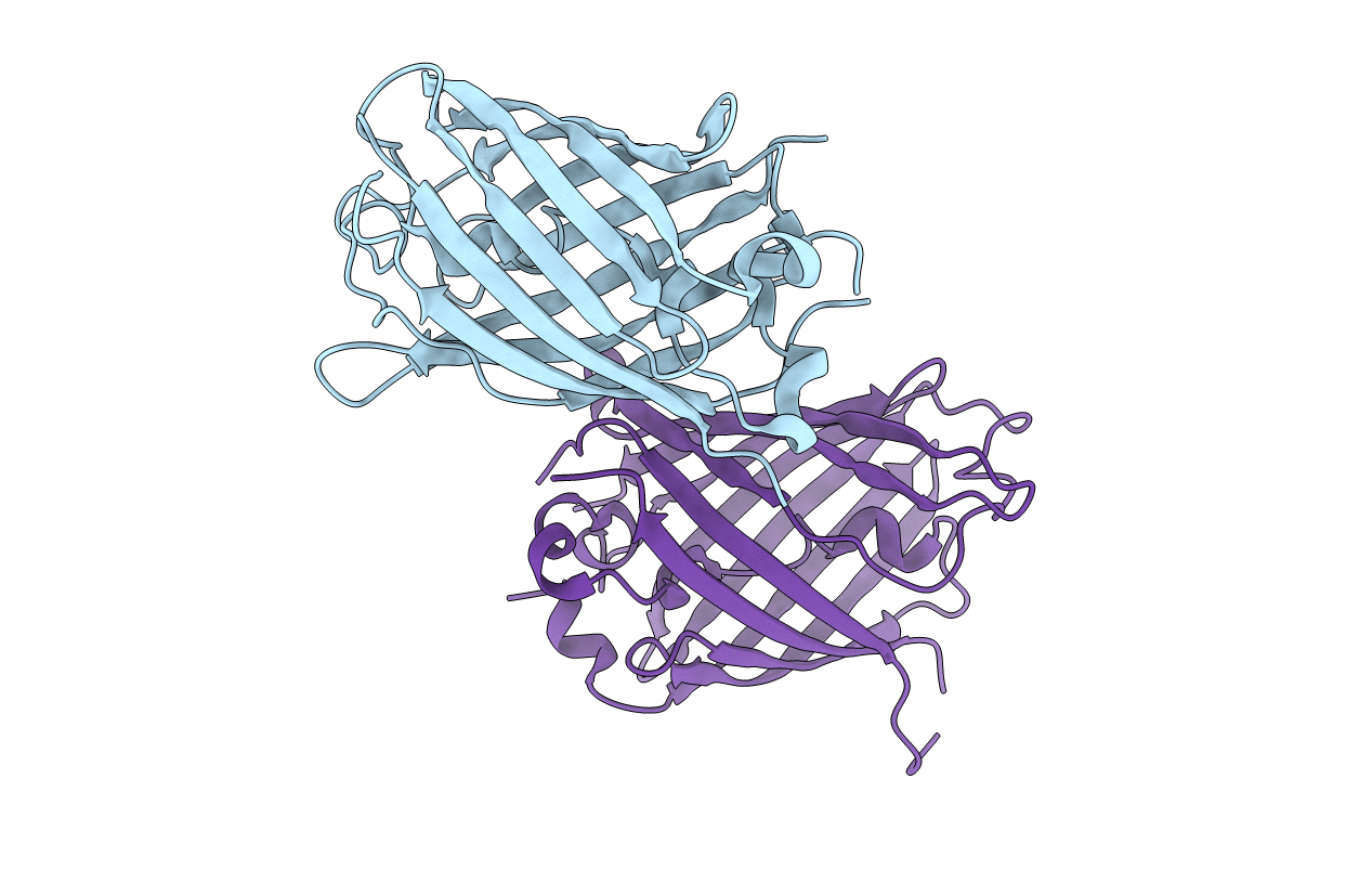
PDB ID:
6OFO
Keywords:
Title:
Crystal structure of split green fluorescent protein (GFP); s10 circular permutant (194-195)
Biological Source:
Source Organism(s):
Aequorea victoria (Taxon ID: 6100)
Expression System(s):
Method Details:
Experimental Method:
Resolution:
2.60 Å
R-Value Free:
0.23
R-Value Work:
0.20
R-Value Observed:
0.20
Space Group:
P 1 21 1